Atopic dermatitis
Atopic dermatitis
Updated: 04/01/2025
© Jun Wang, MD, PhD
General features
- Usually starts in early infancy
- Higher risk to develop other allergic disorders later in life
- More common in women, Asians, Blacks, immigrants from developing countries into developed countries
- Patients tend to have hypersensitive skin
- Usually with family history of atopic dermatitis
- Intermittent course with flares and remissions occurring, for unexplained reasons
- Diagnosis based on clinical features, IgE reactivity and history of allergies
Pathogenesis
- Most likely a combination of skin barrier defects and abnormal immune response
- Filaggrin or SPINK5 mutation: causing increased epidermal permiability
- IgE sensitization
- Imbalance between T cell subgroups
- Cutaneous hyper-reactivity to environmental triggers
- Skin barrier dysfunction worsened by inflammation
Clinical features
- Incessant pruritus
- Relatively age specific site of involvement
Early childhood: Face, extensor sides of extremities,
Older children or adults: Flexural surfaces, side of neck,
popliteal and antecubital fossa
- Primary physical findings including:
Xerosis: Dry skin
Lichenification:
Thickening and hardening of skin
Eczematous lesions
Infant atopic
dermatitis
- Shortly after birth
- Earliest presentation include erythema and exudation
- Start from creases, then cheeks, forehead and scalp, and the extensors of the legs
- Nose and diaper areas usually spared
- Erythematous, ill-defined scaly and crusted patches and plaques
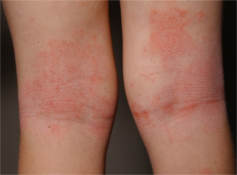
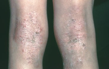
Childhood atopic
dermatitis
- Generalized xerosis
- Flaky and rough skin
- Commonly over the folds, bony protuberances, and forehead
- Eczematous, exudative, scaly and crusted lesions
- Lichenification
Adult atopic
dermatitis
Kaposi varicelliform
eruption
- AKA Eczema herpeticum
- Cutaneous eruption caused by herpes simplex virus, coxsackievirus A16, or vaccinia virus
- Infects preexisting dermatosis
- Most commonly disseminated HSV infection in patients with atopic dermatitis
- Associated with impaired immune function and mechanical barrier defect of affected skin
- Clinical features
Sudden eruption of painful;
edematous; crusted/hemorrhagic vesicles, pustules, or erosions
Areas of the preexisting
dermatosis
- Treated with antivirals
Pathological findings
- Acute to chronic spongiotic dermatitis
- Acute phase: Spongiosis, parakeratosis, lymphocytic exocytosis
- Chronic phase: Epidermal hyperplasia
Diagnosis
- Essential features: must present
- Chronic or relapsing history
- Eczema
- Pruritus
- Morphology and age-specific patterns
- Import features: supportive, see in most cases
- Personal or family history of atopy
- Early onset
- IgE reactivity
- Xerosis
- Associated features: non-specific
- Atypical vascular responses
- Keratosis pilaris, pityriasis alba, etc
- Ocular or periorbital changes
- Perifollicualr accentuation, lichenification, etc
- Moisturization
- Topical steroid
- Immunomodulators
Back to contents
Comments
Post a Comment