Posts

Showing posts with the label respiratory

Practice question III answers Lung and pleural tumors

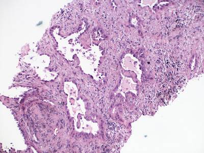
Practice question III answers Lung and pleural tumors © Jun Wang, MD, PhD   1. B. Malignant tumors composed of irregular glands lined by atypical cells are adenocarcinoma s . Abscess is characterized by collection of neutrophils and other inflammatory cells in a necrotic background, but do not have glandular growth. Metastatic renal cell carcinoma does not have glandular pattern and is negative for TTF-1. Small cell carcinoma has solid nests of cells with round to oval molding nuclei and scant cytoplasm. Squamous cell carcinoma has squamous differentiation characterized by intercellular bridges and keratin pearls . 2. D. TTF-1 is usually positive in adenocarcinomas of lung. Calretinin and WT-1 are positive for mesothelioma . Chromogranin is a neuroendocrine marker that is expressed in neuroendocrine tumors, including carcinoid , small cell carcinoma , and olfactory neuroblastoma . P63 is a marker for squamous cell differentiation and is positive for squamous cell carc...

Practice questions III Lung and pleural tumors

Image
Practice questions III Lung and pleural tumors © Jun Wang, MD, PhD   1. Use this case and this image for next three questions . A 75-year-old woman presents with non-productive cough and a 15 lb weight loss in the past 6 months. She does not have fever or chest pain. She has a history of clear cell type renal cell carcinoma at age 57 and was treated with left nephrectomy. Her past medical history is otherwise unremarkable. She does not smoke cigarette nor drink alcohol. Radiologic examination reveals a 2.5 cm lesion in the right middle lobe. An image of the biopsy is shown. There are foci of necrosis with neutrophils. What is most likely the diagnosis?     ( Image credit: Yale Rosen from USA, CC BY-SA 2.0 <https://creativecommons.org/licenses/by-sa/2.0>, via Wikimedia Commons)   A. Abscess B. Adenocarcinoma C. Metastatic renal cell carcinoma D. Small cell carcinoma E. Squamous cell carcinoma   2. A 75-year-old woman presents with non-productive c...

Lymphangioleiomyomatosis

Lymphangioleiomyomatosis Updated: 11/01/2021 © Jun Wang, MD, PhD   General features Cystic destruction with progressive dysfunction of lung Proliferation of lymphatic smooth muscle-like cells Probably originated from perivascular epithelioid cell Most common in reproductive age women Associated with tuberous sclerosis Key clinical features Most common presentations: Dyspnea, cough, pleural effusion, etc Pneumothorax commonly seen Key morphological features Cystic changes Proliferation of spindle cells (LAM cells), may be nodular Genetic abnormalities TSC1 and TSC2 Diagnosis Radiologic studies: Cystic changes Pathological identification of LAM cells Markers Positive: HMB45 Treatment Treatments of complications Lung function improvement Lung transplantation   Back to respiratory tract neoplasms Back to contents  

Practice question answers Lung and pleural tumors

Practice question answers Lung and pleural tumors © Jun Wang, MD, PhD 1. E. Presence of intercellular bridges is consistent with squamous cell differentiation. Irregular contours of the nests and marked pleomorphism are consistent with malignancy. Malignant tumor with squamous cell differentiation in lung is squamous cell carcinoma . Abscess is by definition a collection of pus, but should not have cytological atypia. Adenocarcinomas, including those from lung and colon, have glandular differentiation but not intercellular bridges . Pulmonary small cell carcinoma has solid nests of cells with round to oval molding nuclei and scant cytoplasm. 2. D. p63 is marker for squamous cell differentiation. Calretinin is positive for mesothelioma . Chromogranin is a neuroendocrine marker that is expressed in neuroendocrine tumors, including carcinoid , small cell carcinoma , and olfactory neuroblastoma . CK20 is usually positive for colon adenocarcinoma. TTF-1 is usually positi...

Practice questions Lung and pleural tumors

Practice questions II, Lung and pleural tumors © Jun Wang, MD, PhD 1. Use this case for next two questions . A 69-year-old woman presents with non-productive cough for 3 months. She has a history of stage II sigmoid colon cancer 5 month ago, and was treated with surgery and chemotherapy. She just started a cycle of chemotherapy 1 week ago. She has a 30 year 1 pack per day smoking history. She does not drink alcohol. Radiologic examination reveal a 1.5 cm lesion at the right hilum. Biopsy of the lesion reveal cords and nests with irregular contours composed of cells with large amount eosinophilic cytoplasm and markedly atypical nuclei. Intercellular bridges and squamous pearls are seen. There are foci of necrosis with neutrophils. What is most likely the diagnosis? A. Abscess B. Adenocarcinoma C. Metastatic colon cancer D. Small cell carcinoma E. Squamous cell carcinoma 2. A 69-year-old woman presents with non-productive cough for 3 months. She has a history of st...