Posts

Showing posts with the label esophagus

Practice question answers esophageal pathology

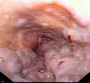
Practice question answers Esophageal pathology Updated: 03/07/2021 © Jun Wang, MD, PhD 1. D. Dysphagia may be caused by abnormal esophageal motility or physical obstructions, including web and ring , as well as cancers . In this case, physical obstruction is ruled out by endoscopy and image studies. High-resolution manometry is used to monitor the pattern of esophageal motility. Chest pain without other presentations is less likely caused by coronary artery or mediastinal abnormality. Biopsy is used when there is suspicious lesions, or symptoms of reflux. 2. C. Diffuse uncoordinated muscular contraction involving entire esophagus is consistent with diffuse esophageal spasm . Achalasia is characterized by lack of progressive peristalsis and partial/incomplete relaxation of lower esophageal sphincter . Esophagitis , including candidiasis and esophageal web have changes can be identified by endoscope or image studies. 3. A. Achalasia is characterized by lack of progres...
Practice questions Esophageal pathology

Image
Practice questions Esophageal pathology Updated: 02/28/2019 © Jun Wang, MD, PhD 1. Use this case for the next two questions . A 35-year-old woman presents with intermittent dysphagia and chest pain for 1 year. The pain is triggered by swallowing large amount of water. It is a sharp pain in the lower substernal area. She denies other symptoms. She has a history of type 2 diabetes but denies any cardiovascular system disorders. She does not smoke cigarette nor drink alcohol. Physical examination reveals no significant abnormalities. Laboratory tests are within normal range. Upper endoscopic exam, 24 hour esophageal impedance-pH monitoring and barium esophagogram reveal no esophageal or stomach abnormalities. What test is appropriate next? A. Coronary angiogram B. EKG C. Esophageal biopsy D. High-resolution manometry E. Sonographic exam for mediastinal abnormalities 2. A 35-year-old woman presents with intermittent dysphagia and chest pain for 1 year. The pain is tr...

Esophageal varices

Esophageal varices   Updated: 02/12/2021 © Jun Wang, MD, PhD General features Dilated tortuous vessels, usually submucosal Common in cirrhotic patients, usually due to alcoholism Etiology Portal hypertension induced collateral between portal and caval systems Clinical presentations Potentially fatal massive hemorrhage if ruptured Dilated submucosal vein in distal esophagus Diagnosis Endoscopy Image studies Treatment Prevent bleeding Treatment of bleeding Back to esophagus pathology Back to contents

Esophageal spasm

Esophageal spasm   Updated: 02/12/2021 © Jun Wang, MD, PhD General features More common in white, women Symptoms may improve over time Nutcracker esophagus/hypertensive peristalsis High-amplitude Coordinated contractions of the distal esophagus Diffuse esophageal spasm Normal amplitude Uncoordinated, simultaneous, or rapidly propagated, contractions of the distal esophageal smooth muscle Clinical presentations Dysphagia, regurgitation, and noncardiac chest pain Diagnosis Best diagnostic modality: High-resolution manometry Treatment Calcium channel blockers, botulinum toxin, nitrates, tricyclic antidepressants, sildenafil, dilatation Myotomy, and esophagectomy Back to esophagus pathology Back to contents

Mallory-Weiss tear

Mallory-Weiss tear Updated: 01/19/2024 © Jun Wang, MD, PhD General features Longitudinal laceration of GE junction or proximal gastric mucosa Associated with alcoholism, hyperemesis gravidarum, occasionally in weight lifters Etiology Failure of reflex relaxation of gastric outlet preceding an antiperistaltic wave Clinical presentations Upper GI bleeds, may cause sudden death Diagnosis Upper endoscopy Treatment Support, vasoconstrictors, transfusions, occasionally balloon tamponade, surgical repair if possible Back to esophagus pathology Back to contents

Esophagus web and ring

Esophagus web and ring   Updated: 02/12/2021 © Jun Wang, MD, PhD Esophagus web Uncommon, semi circumferential mucosal protrusions Most commonly in upper esophagus in women over 40 Episodic dysphagia associated with "bolting" solid food Vascularized fibrous tissue covered by squamous mucosa Treatment: myomectomy, dilation Plummer-Vinson syndrome : Triad of dysphagia , iron-deficiency anemia , and esophagus web , higher risk for esophagus squamous cell carcinoma Esophagus ring Concentric, smooth protrusions of normal esophageal tissue (mucosa, submucosa and muscularis propria) into the lumen Usually in distal esophagus Congenital or scar from caustic liquids Type A: Lower muscular ring; thickened circular smooth muscle with overlying squamous mucosa Type B: Lower mucosal ring/Schatzki’s ring; lined by squamous epithelium and columnar-type epithelium; ring contains connective tissue and fibers of muscularis mucosae Back to esophagus pathology...

Esophagitis

Esophagitis   Updated: 02/12/2021 © Jun Wang, MD, PhD General features Higher incidence in northern Iran and China Classification based on etiology Reflux: Most common Chemical: Alcohol, corrosive acid or alkalis, medical pills Iotrogenic: Chemotherapy, radiation, graft vs host disease Infectious: Virus, bacteria, fungi Part of systemic disease: Bullous pemphigoid, epidermolysis bullosa, Crohn’s, etc Classification based on histological findings Infiltrating inflammatory cells: Eosinophilic, lymphocytic, neutrophilic Inflammatory pattern: Granulomatous, erosive, necrotic, etc Etiology Most common cause: Gastroesophageal reflux ; less commonly infectious Other causes: Alcohol, corrosive acids or alkalis, excessively hot fluids, and heavy smoking, pills Clinical presentations Heartburn (the most common symptom) Regurgitation, upper abdominal discomfort, nausea, bloating, fullness, dysphagia, odynophagia, cough, hoarseness, wheezing, and hematem...

Achalasia

Achalasia   Updated: 02/12/2021 © Jun Wang, MD, PhD General features Lack of progressive peristalsis and partial/incomplete relaxation of lower esophageal sphincter (LES) Preferentially involves circular layer of muscularis propria, which is hypertrophied Markedly higher risk for squamous cell carcinoma Higher risk for aspiration, Barrett's esophagus , Candida infection, gastroesophageal reflux, lower esophageal diverticula, peptic ulceration, stricture Etiology Primary : T cell mediated destruction or complete absence of myenteric ganglion cells in lower third of esophagus Secondary : Allgrove‘s syndrome, amyloidosis, Chagas' disease, diabetic autonomic neuropathy, polio, sarcoidosis, surgical ablation of dorsal motor nuclei, thyroid disease, tumor Clinical presentations Dysphagia (most common), regurgitation, chest pain, heartburn, weight loss Diagnosis Barium swallow: Bird’s beak appearance , esophageal dilatation Esophageal manometry ( standar...

Esophagus cancers

Esophagus cancers   Updated: 02/12/2021 © Jun Wang, MD, PhD General features Squamous cell carcinoma more common worldwide Squamous cell carcinoma : Higher risk in patients with a long history of smoking and alcohol consumption Incidence of adenocarcinoma has increased in developed countries More common in men than in women Most commonly during the sixth and seventh decades of life Risk factors Squamous cell carcinoma : Alcohol and tobacco, caustic injury, betel nut, drinking scalding hot liquid, achalasia , Plummer-Vinson syndrome , etc Adenocarcinoma : Barrett esophagus , reflux esophagitis , obesity, tobacco Clinical presentations Usually asymptomatic at early phase Insidious onset, dysphagia to solids, followed by dysphagia to all food Extreme weight loss Metastasis generally occurs early Masses, with or without ulceration Pathological features Mass, with or without ulcer Squamous cell carcinoma: Cords of atypical cells with squamous different...

Barrett esophagus

Barrett esophagus   Updated: 02/14/2022 © Jun Wang, MD, PhD General features Intestinal metaplasia of distal squamous mucosa Incidence higher in whites, males, obese (especially with central adiposity) May regress after treatment Major risk factor for esophageal adenocarcinoma Etiology Chronic injury, i.e. chronic gastroesophageal reflux  Clinical presentations Long history of heartburn and other reflux symptoms More massive reflux with more numerous and longer episodes than most reflux patients Pathological features Erythematous mucosa Intestinal metaplasia in gastric-type mucosa above the gastroesophageal junction May harbor dysplasia Low grade : Minimal glandular architectural distortion, elongated hyperchromic nuclei in deep glands and surface epithelium High grade : Distorted glandular architecture, markedly atypical nuclei Diagnosis Endoscopic examination with biopsy Treatment No dysplasia: follow up Low grade dysplasia: en...

Pathology of esophagus

Pathology of esophagus Updated: 02/20/19 © Jun Wang, MD, PhD Key anatomy and histological features Muscular tubular structure Striated muscle: upper portion (suprasternal notch and up) Mixed striated and smooth muscle: mid (suprasternal notch to diaphragmatic hiatus): Smooth muscle: lower (diaphragm to GE junction) Squamous epithelium GE junction : Junction of squamous and columnar epithelium Functional disorders Esophageal spasm Achalasia Esophagus web and ring Mallory-Weiss tears Benign esophageal disorders Varices Esophagitis Barrett esophagus Esophageal cancers Practice questions Back to contents