Practice question Acute inflammatory dermatosis
Practice question
Acute inflammatory dermatosis
Updated: 01/08/2020
© Jun Wang, MD, PhD
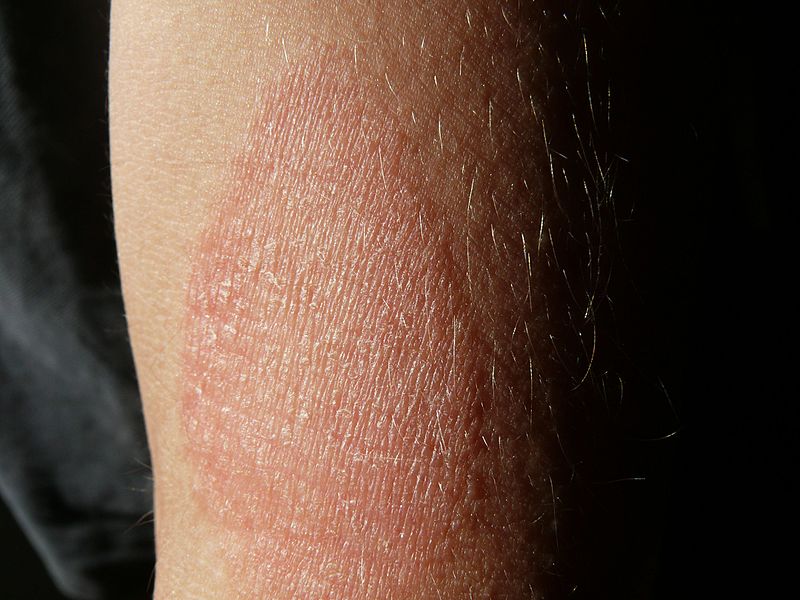
1. Use this
image and this case for the next three questions. A 29-year-old presents
with recurrent elevated skins lesions with itching and burning sensation for 3
months. These lesions are usually at her face, neck, and arms, and last for a
few hours. She denies fever, chills and other constitutional symptoms. Her past
medical history is unremarkable. She does not have any known drug allergy.
Physical examination reveals lesion as shown. What is the most likely
diagnosis?

(Image credit: Enochlau [CC BY-SA 3.0
(http://creativecommons.org/licenses/by-sa/3.0/)])
A. Allergic contact dermatitis
B. Atopic dermatitis
C. Erythema multiformes
D. Guttate psoriasis
E. Urticaria
2. A 29-year-old presents with recurrent elevated
skins lesions with itching and burning sensation for 3 months. These lesions
are usually at her face, neck, and arms, and last for a few hours. She denies
fever, chills and other constitutional symptoms. Her past medical history is
unremarkable. She does not have any known drug allergy. Physical examination
reveals lesion as shown. What is the most likely cause of these findings?
(Image credit: Enochlau [CC BY-SA 3.0 (http://creativecommons.org/licenses/by-sa/3.0/)])
A. Chronic rubbing
B. Desmosome damage
C. Hemidesmosome damage
D. Increased vascular permeability
E. Lymphocytes mediated keratinocyte injury
3. A 29-year-old presents with recurrent elevated
skins lesions with itching and burning sensation for 3 months. These lesions
are usually at her face, neck, and arms, and last for a few hours. She denies
fever, chills and other constitutional symptoms. Her past medical history is
unremarkable. She does not have any known drug allergy. Physical examination reveals
lesion as shown. What cell is likely associated with initiating these symptoms?
(Image credit: Enochlau [CC BY-SA 3.0
(http://creativecommons.org/licenses/by-sa/3.0/)])
A. Eosinophils
B. Fibroblasts
C. Lymphocyte
D. Mast cells
E. Neutrophils
4. Use this
image and this case for the next three questions. A 3-year-old boy presents with fever, fatigue, myalgias, painful oral ulcer and painless reddish
skin lesions at his upper and lower extremities for 2 days. He has had a cold 2
weeks ago. His past medical history include herpes infections. Physical
examination reveals a temperature of 38.5 degree Celsius, heart rate of 110
bpm, blood pressure 115/85 mmHg. Multiple skin lesions are seen as shown in the
image. These skin lesions have a relatively symmetrical distribution, involving
his upper and lower extremities. A few erosions with crusts are seen in the buccal
mucosa. No other abnormalities are noted. What is most likely the diagnosis?

(Photo Credit: Puppy123456 [Public domain])
A. Dermatophytosis
B. Erythema multiforme major
C. Erythema multiforme minor
D. Guttate psoriasis
E. Stevens Johnson syndrome
5. A 3-year-old boy presents with fever, fatigue, myalgias, painful oral ulcer and painless reddish
skin lesions at his upper and lower extremities for 2 days. He has had a cold 2
weeks ago. His past medical history include herpes infections. Physical
examination reveals a temperature of 38.5 degree Celsius, heart rate of 110
bpm, blood pressure 115/85 mmHg. Multiple skin lesions are seen as shown in the
image. These skin lesions have a relatively symmetrical distribution, involving
his upper and lower extremities. A few erosions with crusts are seen in the buccal
mucosa. No other abnormalities are noted. What is most likely cause of these findings?
(Photo Credit: Alborz Fallah [CC BY-SA 3.0 (https://creativecommons.org/licenses/by-sa/3.0)])
A. Autoantibodies against BP180
B. Cornificaiton defects
C. Disseminated fungal infection
D. Sweat gland dysfunction
E. T-cell mediated epidermis damage
6. A 3-year-old boy presents with fever, fatigue, myalgias, painful oral ulcer and painless reddish
skin lesions at his upper and lower extremities for 2 days. He has had a cold 2
weeks ago. His past medical history include herpes infections. Physical
examination reveals a temperature of 38.5 degree Celsius, heart rate of 110
bpm, blood pressure 115/85 mmHg. Multiple skin lesions are seen as shown in the
image. These skin lesions have a relatively symmetrical distribution, involving
his upper and lower extremities. A few erosions with crusts are seen in the buccal
mucosa. No other abnormalities are noted. What is most likely associated with these presentations?
(Photo Credit: Alborz Fallah [CC BY-SA 3.0 (https://creativecommons.org/licenses/by-sa/3.0)])
A. Chemical irritation
B. Drug reaction
C. Fungal infection
D. UV-light damage
E. Viral infection
7. Use this case for the next two questions. A 41-year-old man presents
with fever, diffuse body pain and sloughing of skin, a day after he was treated
with valproic acid for seizure. He had a brain trauma due to automobile accident.
He has shingle and type 2 diabetes. Physical examination reveals diffuse
erythematous changes and erosion involving his oral cavity, eyes, face, trunk
and extremities. There are irregular dull red macules and papules, some have
concentric ring appearance. Blistering changes are seen predominantly at his face
and trunk. The skin sloughing is estimated to be around 5% of total body
surface area. What is most likely the diagnosis?
A. Bullous pemphigoid
B. Erythema multiforme major
C. Erythema multiforme minor
D. Stevens Johnson syndrome
E. Toxic epidermal necrolysis
8. A 41-year-old man presents with fever, diffuse body
pain and sloughing of skin, a day after he was treated with valproic acid for
seizure. He had a brain trauma due to automobile accident. He has shingle and
type 2 diabetes. Physical examination reveals diffuse erythematous changes and
erosion involving his oral cavity, eyes, face, trunk and extremities. There are
irregular dull red macules and papules, some have concentric ring appearance. Blistering
changes are seen predominantly at his face and trunk. The skin sloughing is
estimated to be around 5% of total body surface area. What is most likely associated
with these presentations?
A. Brain trauma
B. Chemical irritation
C. Drug reaction
D. Hemidesmosome demage
E. Viral infection
9. Use this
image and this case for the next two questions. A 35-year-old man presents
with pruritic blisters at his forearms after cleaning his backyard. He denies
other symptoms. His past medical history is unremarkable. Physical examination
reveals blisters up to 2.5 cm in greatest dimension in his forearms as shown in
the image. Biopsy of the lesions reveal marked spongiosis with intraepidermal
vesicles, and diffuse lymphocytic and eosinophilic infiltrate. No acantholysis
is seen. What is most likely the diagnosis?

(Image credit: Joelloughead [Public domain])
A. Allergic contact dermatitis
B. Bullous pemphigoid
C. Dermatitis herpetiformis
D. Lichen planus, bullous type
E. Pemphigus vulgaris
10. A 35-year-old man presents with pruritic blisters
at his forearms after cleaning his backyard. He denies other symptoms. His past
medical history is unremarkable. Physical examination reveals blisters up to
2.5 cm in greatest dimension in his forearms as shown in the image. Biopsy of
the lesions reveal marked spongiosis with intraepidermal vesicles, and diffuse
lymphocytic and eosinophilic infiltrate. No acantholysis is seen. What is the
cause of these findings?
(Image credit: Joelloughead [Public domain])
A. Autoantibodies against desmoglein 1 or 3
B. Autoantibodies against BP180 or BP230
C. Fungal infection
D. IgM containing immunocomplex deposit at
dermoepidermal junction
E. T-cell mediated keratinocytes injury
11. Use this
image and this case for the next three questions. A 2-month-old boy
presents with itchy skin rash on his face and chest. He was born full term with
unremarkable prenatal and delivery courses. Physical examination reveals scaly
rash as shown in the image. The rash involves majority of the face but not the
nose. No other abnormalities are noted. What is likely the diagnosis?

(Image credit: Gzzz [CC BY-SA 4.0
(https://creativecommons.org/licenses/by-sa/4.0)])
A. Allergic contact dermatitis
B. Atopic dermatitis
C. Dermatitis herpetiformis
D. Pemphigus erythematosus
E. Systemic lupus erythematosus
12. A 2-month-old boy presents with itchy skin rash on
his face and chest. He was born full term with unremarkable prenatal and
delivery courses. Physical examination reveals scaly rash as shown in the
image. The rash involves majority of the face but not the nose. No other
abnormalities are noted. What is the cause of these presentations?
(Image credit: Gzzz [CC BY-SA 4.0
(https://creativecommons.org/licenses/by-sa/4.0)])
A. Autoantibody against phospholipid
B. Cornification defect
C. Drug reaction
D. Fungal infection
E. Skin barrier defect
13. A 2-month-old boy presents with itchy skin rash on
his face and chest. He was born full term with unremarkable prenatal and
delivery courses. Physical examination reveals scaly rash as shown in the
image. The rash involves majority of the face but not the nose. No other
abnormalities are noted. What disorder he likely to develop in the future?
(Image credit: Gzzz [CC BY-SA 4.0
(https://creativecommons.org/licenses/by-sa/4.0)])
A. Allergic disorders
B. Basal cell carcinoma
C. Infections
D. Malabsorption
E. Squamous cell carcinoma
14. Use this
image and this case for the next three questions. A 35-year-old woman
presents with an intermittent pruritic skin lesion at her antecubital fossae for
2 months. She does not have other symptoms. She has a history of asthma since
age 8. Physical examination is unremarkable except the skin lesion shown in the
image. Per palpation, this lesion has a rubbery consistency. Biopsy of the
lesion reveals hyperkeratosis, parakeratosis, epidermal hyperplasia with elongated
rete ridges and mild spongiosis. Special stains are negative for fungal hyphae.
What is the diagnosis?
(Image credit: G.steph.rocket [CC BY-SA 3.0 (https://creativecommons.org/licenses/by-sa/3.0)])
A. Allergic contact dermatitis
B. Atopic dermatitis
C. Dermatophytosis
D. Lichen planus
E. Psoriasis
15. A 35-year-old woman presents with an intermittent pruritic
skin lesion at her antecubital fossae for 2 months. She does not have other
symptoms. She has a history of asthma since age 8. Physical examination is
unremarkable except the skin lesion shown in the image. Per palpation, this
lesion has a rubbery consistency. Biopsy of the lesion reveals hyperkeratosis, parakeratosis,
epidermal hyperplasia with elongated rete ridges and mild spongiosis. Special
stains are negative for fungal hyphae. What blood test is likely to be abnormal?
(Image credit: G.steph.rocket [CC BY-SA 3.0 (https://creativecommons.org/licenses/by-sa/3.0)])
A. Absolute lymphocytes
B. Absolute neutrophils
C. IgA
D. IgE
E. IgG
16. A 35-year-old woman presents with an intermittent pruritic skin lesion at her antecubital fossae for 2 months. She does not have other symptoms. She has a history of asthma since age 8. Physical examination is unremarkable except the skin lesion shown in the image. Per palpation, this lesion has a rubbery consistency. Biopsy of the lesion reveals hyperkeratosis, parakeratosis, epidermal hyperplasia with elongated rete ridges and mild spongiosis. Special stains are negative for fungal hyphae. What is the cause of skin thickening of this lesion?
(Image credit: G.steph.rocket [CC BY-SA 3.0 (https://creativecommons.org/licenses/by-sa/3.0)])
A. Chronic rubbing
B. Fungal infection
C. Lymphocytic infiltrate
D. Sweat gland dysfunction
E. Vascular hyperpermiability
E. Vascular hyperpermiability
17. Use this
image for this question. A 7-year-old boy presents with sudden onset of
painful vesicles on his face for a day. He has a history of atopic dermatitis
and is treated with topical steroids. He does not have fever or other symptoms.
Physical examination reveals numerous small vesicles with purulent contents,
some have ruptures and are covered yellowish crust. Biopsy reveals findings as
shown in the image. What is the diagnosis?

(Image credit: Yale Rosen from USA [CC BY-SA 2.0
(https://creativecommons.org/licenses/by-sa/2.0)])
A. Dermatitis herpetiformis
B. Irritant contact dermatitis
C. Kaposi varicelliform eruption
D. Pustular psoriasis
E. Shingle
18. Use this
image for this question. A 48-year-old man presents with pruritic visicles
at his fingers and palms. He has type 2 diabetes and shingles. Physical
examination reveals clusters of small vesicles with clear contents, as shown in
the image. No other abnormalities are noted. What is most likely the diagnosis?

Image credit (commonswiki)
A. Allergic contact dermatitis
B. Dermatitis herpetiformis
C. Dyshydrotic eczema
D. Pustular psoriasis
E. Shingles
19. A 15-year-old girl presents with a pruritic skin
lesion at the extensor side of her left arm for 6 months. Her past medical
history is unremarkable. Physical examination reveals a 1.5 oval shaped plaque
with erythematous base and dry, cracked surface with mild scaling at both
center and periphery. No other abnormality is noted. What is the diagnosis?
A. Dermatophytosis
B. Dyshidrotic eczema
C. Lichen planus
D. Nummular eczema
E. Psoriasis
20. A 21-year-old man presents with new pruritic
rashes at his left leg. He had seborrheic dermatitis 2 weeks ago and was
treated with topical steroid. His past medical history is otherwise
unremarkable. Physical examination reveals diffuse erythematous papules with
yellowish scaling on his left leg. Biopsy of these new lesions reveals parakeratosis
and mild spongiosis, that are prominent around hair follicles. No fungal hyphae
are seen per special stain. What is the most likely diagnosis?
A. Dermatitis herpetiformis
B. Dermatophytosis
C. ID reaction
D. Kaposi varicelliform eruption
E. Pustular psoriasis
21. A 77-year-old presents pruritic dry skin in both legs
for 5 years. The symptoms are more prominent in winter months. He has type 2
diabetes and hypertension. His past medical history is otherwise unremarkable. Physical
examination reveals rubbery very dry skin with irregular cracks, fissures and
scaling of both legs. No other abnormalities are noted. What is most likely the
diagnosis?
A. Atopic dermatitis
B. Dermatophytosis
C. Lichen planus
D. Psoriasis
E. Xerotic eczema
22. Use this image for the next two questions. A 45-year-old woman presents to the clinic with persistent redness on her cheeks and nose for the past 6 months. She does not have other symptoms. She has had intermittent episodes of flushing, especially after sunlight exposure for the last five years. Her family history is unremarkable. She does not smoke cigarettes nor drink alcohol. An image of her face is shown. Her laboratory tests are within normal ranges. What is most likely associated with her facial changes?

(Image credit: Michael Sand, Daniel Sand, Christina Thrandorf, Volker Paech, Peter Altmeyer, Falk G Bechara, CC BY 2.5 <https://creativecommons.org/licenses/by/2.5>, via Wikimedia Commons)
A. Antinuclear antibodies
B. Demodex mites
C. Malassezia
D. Myositis-specific autoantibodies
E. UV light
23. A 45-year-old woman presents to the clinic with persistent redness on her cheeks and nose for the past 6 months. She does not have other symptoms. She has had intermittent episodes of flushing, especially after sunlight exposure for the last five years. Her family history is unremarkable. She does not smoke cigarettes nor drink alcohol. An image of her face is shown. Her laboratory tests are within normal ranges. What is the diagnosis?
(Image credit: Michael Sand, Daniel Sand, Christina Thrandorf, Volker Paech, Peter Altmeyer, Falk G Bechara, CC BY 2.5 <https://creativecommons.org/licenses/by/2.5>, via Wikimedia Commons)
A. Cutaneous lupus erythematosus
B. Dermatomyositis
C. Rosacea
D. Seborrheic dermatitis
E. Sun-damaged skin

(Image credit: Michael Sand, Daniel Sand, Christina Thrandorf, Volker Paech, Peter Altmeyer, Falk G Bechara, CC BY 2.5 <https://creativecommons.org/licenses/by/2.5>, via Wikimedia Commons)
A. Antinuclear antibodies
B. Demodex mites
C. Malassezia
D. Myositis-specific autoantibodies
E. UV light
23. A 45-year-old woman presents to the clinic with persistent redness on her cheeks and nose for the past 6 months. She does not have other symptoms. She has had intermittent episodes of flushing, especially after sunlight exposure for the last five years. Her family history is unremarkable. She does not smoke cigarettes nor drink alcohol. An image of her face is shown. Her laboratory tests are within normal ranges. What is the diagnosis?
(Image credit: Michael Sand, Daniel Sand, Christina Thrandorf, Volker Paech, Peter Altmeyer, Falk G Bechara, CC BY 2.5 <https://creativecommons.org/licenses/by/2.5>, via Wikimedia Commons)
A. Cutaneous lupus erythematosus
B. Dermatomyositis
C. Rosacea
D. Seborrheic dermatitis
E. Sun-damaged skin
Back to acute
inflammatory dermatosis
Back to contents
Comments
Post a Comment