alpha-thalassemia
a-thalassemia
Updated: 07/13/2026
© Jun Wang, MD, PhD
General features
- Anemia associated with reduced Hb, hemolysis and ineffective erythropoiesis
- Reduced HbA, and presence of HbH and/or Hb Bart
- DNA analysis of gene copy numbers required for diagnosis
Pathogenesis
- Deletions of one or more a globin gene
- Reduced or absent synthesis of a globin
- Unpaired b chains and g chains (in infants) more soluble and form fairly stable tetramers
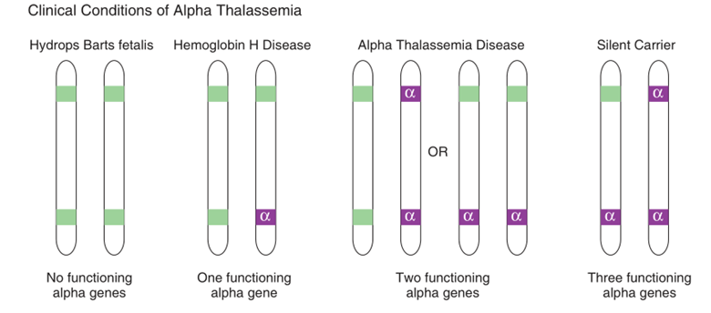
Classification
- Based on number of a globin gene deleted
- Silent carrier state
o Deletion of one a globin gene
o Minimal reduction in a globin synthesis
o Usually asymptomatic
o May have slight microcytosis
o Diagnosed by genetic test
- a thalassemia trait
o Deletion of two a globin genes
o Usually not associated with anemia
o High RBC count with low MCV and MCH
- HbH disease
o Deletion of 3 a globin genes
o HbH (over 30%), composed of 4 b chains
- Alpha thalassemia major (ATM) (Hb Bart fetal hydrops)
o A type of non-immune hydrops fetalis
o Deletion of 4 a globin genes
o Usually died in utero
o Greater than 30% Hb Bart (aka Bart’s Hb, Hb Bart’s), composed of 4 gamma chains
o HbH may present, but no HbA or HbF
a thalassemia trait
- Deletion of two genes
- Cis
o Both genes from the same chromosome
o SEA, THAI, FIL type deletion
o More common in Southeast Asians
o Offspring at higher risk for HbH disease or hydrops fetalis
- Trans
o Deletion involving both chromosomes
- Usually NO anemia
- High RBC count with low MCV and MCH
- Normal Hb electrophoresis
- DNA analysis needed to confirm diagnosis
HbH disease
- Deletion of 3 a genes
- Formation of HbH
o 4 b chains
o ≥ 30% needed for diagnosis
o High oxygen affinity for oxygen
o Tissue hypoxia disproportionate to absolute level of Hb
o Oxidation resulting in formation of intracellular inclusions promoting red cell sequestration and phagocytosis
o Detected by hemoglobin electrophoresis or HPLC
- Poikilocytosis: microcytes, target cells, schistocytes, tear drop cells
- No iron overload
- No extramedullary hematopoiesis
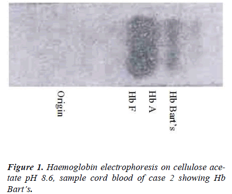
Hb Bart fetal hydrops
- Deletion of 4 a genes
- Formation of Hb Bart
o Tetramers of g globin
o ≥ 30% for diagnosis
o Very high affinity for oxygen and does not dissociate
o Severe tissue hypoxia
o Detected by hemoglobin electrophoresis or HPLC
o May have HbH (up to 30% of total hemoglobin)
- Death in utero unless intrauterine blood transfusions
- Embryonic hemoglobins (Gower I, Portland, no alpha globins) in first trimester
- Hb Bart formed later in pregnancy, causing severe tissue hypoxia
- Tissue anoxia may lead to fetal death
- If fetal transfusions given and infant born alive, lifelong supportive transfusions necessary unless bone marrow/stem cell transplant
Hb electrophoresis findings
- Carrier state: Normal
- Trait: mild decrease (~10-15%) of HbA, no abnormal Hb
- HbH
o Combination of HbA, HbH, and Hb Bart
o HbH ≥ 30% for diagnosis
- Hydrops fetalis:
o NO HbA or HbF
Management
- Mild forms: NO specific treatment, iron supplementation
- HbH
o Supplementation of folic acid
o Lifelong transfusion likely for severe anemia
- Hb Bart
Comments
Post a Comment