Practice question Pathology of breast III
Practice question
Pathology of breast III
© Jun Wang, MD, PhD
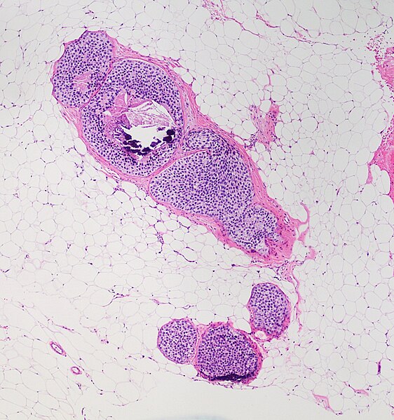
1. Use this image for the next two questions. A 42-year-old woman presents to the clinic with a left breast mass for 1 month. She does not have history of trauma. She has had type 2 diabetes for 5 years and has been treated with metformin. Her family history is significant for multiple cancers including endometrial carcinoma and benign ovarian tumors. Physical examination reveals a 2.5 cm mobile firm mass at the outer upper quadrant of her left breast. No other abnormalities are noted. An image of her biopsy is shown. What is the diagnosis?
.jpg)
(No machine-readable author provided. KGH assumed (based on copyright claims)., CC BY-SA 3.0 <http://creativecommons.org/licenses/by-sa/3.0/>, via Wikimedia Commons)
A. Diabetic mastopathy
B. Fibroadenoma
C. Fibrocystic changes
D. Invasive ductal carcinoma
E. Invasive lobular carcinoma
F. Usual ductal hyperplasia
2. A 42-year-old woman presents to the clinic with a left breast mass for 1 month. She does not have history of trauma. She has had type 2 diabetes for 5 years and has been treated with metformin. Her family history is significant for multiple cancers including endometrial carcinoma and benign ovarian tumors. Physical examination reveals a 2.5 cm mobile firm mass at the outer upper quadrant of her left breast. No other abnormalities are noted. An image of her biopsy is shown. What is the diagnosis?
What will be the most likely immunohistochemistry study finding?
(No machine-readable author provided. KGH assumed (based on copyright claims)., CC BY-SA 3.0 <http://creativecommons.org/licenses/by-sa/3.0/>, via Wikimedia Commons)
B. Loss of E-cadherin
C. p53 reactivity in glandular cells
D. p63 positive cells at base of ducts
E. Polyclonal B cells
3. Use this image for the next two questions. A 43-year-old woman presents with microcalcifications identified by mammogram screening. Her past medical history is unremarkable. Physical examination reveals no abnormalities. An image of her biopsy is shown. What is the diagnosis?

(Image credit: Manuel Medina from Osuna, España, Public domain, via Wikimedia Commons)
B. Ductal carcinoma in situ
C. Fibrocystic changes
D. Invasive ductal carcinoma
E. Mucinous carcinoma
4. A 43-year-old woman presents with microcalcifications identified by mammogram screening. Her past medical history is unremarkable. Physical examination reveals no abnormalities. An image of her biopsy is shown. What is most likely associated with these changes?
(Image credit: Manuel Medina from Osuna, España, Public domain, via Wikimedia Commons)
B. Estrogen effect
C. Her2 amplification
D. Loss of E-cadherin
E. Recent trauma
5. Use this image for the next question. A 51-year-old woman presents to the clinic with a slowly growing right breast mass for 2 months. She denies other symptoms. She has a family history of breast and ovarian cancers in the maternal side. Physical examination reveals a 2.5 cm mobile mass at the outer upper quadrant. An image of her biopsy is shown. What is the diagnosis?

B. Ductal carcinoma in situ
C. Invasive ductal carcinoma
D. Papilloma
E. Phyllodes tumor
6. Use this image for the next two questions. A 44-year-old woman presents to the clinic for a mammographic finding of left breast retroareolar growth. She has a history of diabetes. Her family history reveals multiple members with colon and lung cancers. Physical examination reveals no significant abnormality. An image of her biopsy is shown. No significant cytological atypia is seen. CD10 stain reveals positive reactivity at the base of epithelial lining. What is the diagnosis?

(Image: Ed Uthman from Houston, TX, USA, CC BY 2.0 <https://creativecommons.org/licenses/by/2.0>, via Wikimedia Commons)
C. Intraductal papilloma
D. Invasive ductal carcinoma
E. Lobular carcinoma in situ
7. A 44-year-old woman presents to the clinic for a mammographic finding of left breast retroareolar growth. She has a history of diabetes. Her family history reveals multiple members with colon and lung cancers. Physical examination no significant abnormality. An image of her biopsy is shown. No significant cytological atypia is seen. CD10 stain reveals positive reactivity at the base of epithelial lining. What is the most common clinical presentation of her condition?
(Image: Ed Uthman from Houston, TX, USA, CC BY 2.0 <https://creativecommons.org/licenses/by/2.0>, via Wikimedia Commons)
B. Eczematous nipple changes
C. Mammographic finding of microcalcification
D. Multiple nodular growth
E. Palpable mass
8. Use this image for the next question. A 44-year-old woman presents to the clinic for a mammographic finding of left breast architecture changes. Her past medical history is unremarkable. Physical examination revelas no significant abnormality. Image study reveals a 0.7 cm poorly demarcated mass. An image of her biopsy is shown. No significant cytological atypia is seen. CD10 stain reveals intake myoepithelial layer. What is the diagnosis?

(Image: Mikael Häggström, M.D. Author info - Reusing images- Conflicts of interest: NoneMikael Häggström, M.D. Consent note: Consent from the patient or patient's relatives is regarded as redundant, because of absence of identifiable features (List of HIPAA identifiers) in the media and case information (See also HIPAA case reports guidance)., CC0, via Wikimedia Commons)
B. Intraductal papilloma
C. Invasive ductal carcinoma
D. Radial scar
E. Sclerosing adenosis
9. Use this image for the next question. A 44-year-old woman presents to the clinic for a mammographic finding of left breast architecture changes. Her past medical history is unremarkable. Physical examination revelas no significant abnormality. An image of her biopsy is shown. No significant cytological atypia is seen. CD10 stain reveals intake myoepithelial layer. What is the diagnosis?

B. Intraductal papilloma
C. Invasive ductal carcinoma
D. Radial scar
E. Sclerosing adenosis
10. Use this image for the
next two questions. A 44-year-old woman presents with microcalcifications and
altered architecture identified by mammogram screening. Her past medical
history is unremarkable. Physical examination reveals no abnormalities. An
image of her biopsy is shown. CD10 stain reveals intake myoepithelial layer. What
immunohistochemistry marker should be examined next?
(Image: Ed Uthman from Houston, TX, USA, CC BY 2.0 <https://creativecommons.org/licenses/by/2.0>, via Wikimedia Commons)
B. Estrogen receptor
C. Her2
D. p53
E. Progesterone receptor
11. A 44-year-old woman presents with microcalcifications and altered architecture identified by mammogram screening. Her past medical history is unremarkable. Physical examination reveals no abnormalities. An image of her biopsy is shown. CD10 stain reveals intake myoepithelial layer.
These cells are positive for E-cadherin. What is the diagnosis?
A. Ductal carcinoma in situ
B. Intraductal papilloma
C. Invasive ductal carcinoma
D. Radial scar
E. Sclerosing adenosis
.jpg)
(Image: No machine-readable author provided. KGH assumed (based on copyright claims)., CC BY-SA 3.0 <http://creativecommons.org/licenses/by-sa/3.0/>, via Wikimedia Commons)
B. Intraductal papilloma
C. Invasive ductal carcinoma
D. Radial scar
E. Sclerosing adenosis

(Image: Difu Wu., CC BY-SA 3.0 <https://creativecommons.org/licenses/by-sa/3.0>, via Wikimedia Commons)
B. Estrogen receptor
C. Her2
D. p53
E. Progesterone receptor
14. A 44-year-old woman presents with microcalcifications and altered architecture identified by mammogram screening. Her past medical history is unremarkable. Physical examination reveals no abnormalities. An image of her biopsy is shown. CD10 stain reveals intake myoepithelial layer.
These cells are negative for E-cadherin. What immunohistochemistry marker should be examined next?
B. Invasive ductal carcinoma
C. Invasive lobular carcinoma
D. Lobular carcinoma in situ
E. Usual ductal hyperplasia

(Image: Calicut Medical College, CC BY-SA 4.0 <https://creativecommons.org/licenses/by-sa/4.0>, via Wikimedia Commons)
A. CK5/6
B. E-cadherin
C. Her2
D. p53
(Image: Calicut Medical College, CC BY-SA 4.0 <https://creativecommons.org/licenses/by-sa/4.0>, via Wikimedia Commons)
B. Invasive ductal carcinoma
C. Invasive lobular carcinoma
D. Lobular carcinoma in situ
E. Usual ductal hyperplasia

(Image: Mikael Häggström, M.D. Author info - Reusing images- Conflicts of interest: NoneMikael Häggström, M.D. Consent note: Consent from the patient or patient's relatives is regarded as redundant, because of absence of identifiable features (List of HIPAA identifiers) in the media and case information (See also HIPAA case reports guidance)., CC0, via Wikimedia Commons)
B. ER negative, Her2 positive
C. ER positive, Her2 negative
D. ER positive, Her2 positive
Additional immunohistochemistry studies reveals these epithelial cells are negative for estrogen receptor, progesterone receptor and Her2, in a background of mixed CD3+ and CD20+ lymphocytes. What is the diagnosis?
(Image: Mikael Häggström, M.D. Author info - Reusing images- Conflicts of interest: NoneMikael Häggström, M.D. Consent note: Consent from the patient or patient's relatives is regarded as redundant, because of absence of identifiable features (List of HIPAA identifiers) in the media and case information (See also HIPAA case reports guidance)., CC0, via Wikimedia Commons)
A. Diffuse large B cell lymphoma
B. Ductal carcinoma in situ
C. Invasive ductal carcinoma
D. Invasive lobular carcinoma
E. Medullary carcinoma
F. Metastatic lung adenocarcinoma
19. A 51-year-old woman presents with left breast mass for a month. She has a history of lung adenocarcinoma 5 years ago that was treated with surgery and chemotherapy. She has 30 pack year history of cigarette smoking but has quit since she was diagnosed with lung cancer. Physical examination reveals a firm fixed mass at the lower inner quadrant. An image of her biopsy is shown. Per immunohistochemistry studies, these cells are positive for E-cadherin and negative for TTF-1.
Additional immunohistochemistry studies reveals these epithelial cells are negative for estrogen receptor, progesterone receptor and Her2, in a background of mixed CD3+ and CD20+ lymphocytes. What is genetic abnormality is most likely associated with this lesion?
A. Bcl2
B. BRCA1
C. CDH1
D. CHEK2
E. Cyclin D1

(Image: Ed Uthman from Houston, TX, USA, CC BY 2.0 <https://creativecommons.org/licenses/by/2.0>, via Wikimedia Commons)
B. Invasive ductal carcinoma
C. Invasive lobular carcinoma
D. Mucinous carcinoma
E. Small lymphocytic lymphoma
(Image: Ed Uthman from Houston, TX, USA, CC BY 2.0 <https://creativecommons.org/licenses/by/2.0>, via Wikimedia Commons)
B. BRCA1
C. CDH1
D. Her2
E. MIR15A/MIR16A
 (Sarahkayb,
CC BY-SA 4.0 <https://creativecommons.org/licenses/by-sa/4.0>, via
Wikimedia Commons)
(Sarahkayb,
CC BY-SA 4.0 <https://creativecommons.org/licenses/by-sa/4.0>, via
Wikimedia Commons)B. Invasive ductal carcinoma
C. Invasive lobular carcinoma
D. Junctional nevus
E. Melanoma
F. Paget disease
(Sarahkayb, CC BY-SA 4.0 <https://creativecommons.org/licenses/by-sa/4.0>, via Wikimedia Commons)
A. Ductal carcinoma
B. Ductal ectasia
C. Fibrocystic changes
D. Lobular carcinoma
E. Usual ductal hyperplasia
Comments
Post a Comment