Posts

Seborrheic keratosis

Seborrheic keratosis Updated: 03/27/2019 © Jun Wang, MD, PhD General features Benign epidermal proliferation Most common benign tumor in elder population Unclear etiology Less common in people with darker skin May harbor FGFR3 mutation Squamous cell carcinoma or melanoma may arise Clinical features Sharply demarcated pigmented Soft, tan-black, "greasy" surface Commonly on trunk Can occur anywhere except palms and soles Leser-TrĂ©lat sign Sudden appearance or increase in number and size of seborrheic keratosis Associated with internal malignancy Probably paraneoplastic phenomenon More commonly associated with GI malignancy Pathological features Basaloid cell proliferation Pseudohorn cyst No atypia May be pigmented Management Removal Back to skin tumors Back to contents
Pilomatricoma

Pilomatricoma Updated: 03/28/2019 © Jun Wang, MD, PhD General features AKA pilomatrixoma, calcified epithelioma of Malherbe Most benign  Arising from hair matrix Usually children and young adults in head, neck or upper extremities Likely associated with CTNNB (beta-catenin) mutation or bcl-2 overexpression Aggressive pilomatrixoma : Atypical histology, locally invasive with local recurrence Malignant pilomatrixoma : Cytologic atypia, infiltrative border, transition to squamous cells, necrosis, local recurrence and metastasize Clinical features Solitary firm nodule Pathological features Solid nests of basaloid cells Abrupt keratinization Ghost cells , often foreign body reaction, calcification or ossification Management Excision Back to skin tumors Back to skin pathology Back to contents

Pilar cyst

Pilar cyst   Updated: 03/02/2021 © Jun Wang, MD, PhD General features AKA trichilemmal cyst, isthmus-catagen cyst or a "wen" Most commonly seen in scalp More common in women May be familial with autosomal dominant inheritance Probably originated from root sheath of hair follicle Clinical features Slow growing Firm swelling May be ruptured and inflamed Easy to remove Pathological features Cysts lined by squamous epithelium without granular layer Filled with homogenous material   Management Excision Back to skin tumors Back to skin pathology Back to contents

Merkel cell carcinoma of skin

Merkel cell carcinoma of skin   Updated: 03/17/2021 © Jun Wang, MD, PhD General features Uncommon Aggressive neuroendocrine tumor Origin and etiology unclear Commonly in the head and neck, elderly and light skin population May be associated with Merkel cell polyomavirus, UV, radiation, and immunosuppresion Tendency to recur and metastasize Distant metastasis nearly fatal Clinical features Painless tumor Rapid growing Biopsy needed for diagnosis Key pathological features Sheets of poorly differentiated cells Monotonous round tumor cells with scant cytoplasm Diffuse infiltration of subcutis Markers Positive: neuroendocrine markers (CD56, chromogranin, synaptophysin, etc), CK20 Negative: CD45 (rule out lymphoma ), TTF1 (rule out metastatic pulmonary small cell carcinoma ) Management Excision Radiation Chemotherapy Back to skin tumors Back to skin pathology Back to contents

Melanoma

Image
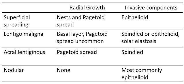
Melanoma   Updated: 03/10/2023 © Jun Wang, MD, PhD General features Malignancy of melanocytes, predominantly in skin May involve eyes, ears, GI tract, leptomeninges, mucous membranes Majority of skin cancer deaths More common in older population Strong association with UV light (sun or artificial) exposure Melanoma in situ: Atypical melanocytes limited in epidermis Invasive melanoma: Atypical melanocytes invade into dermis and beyond Prognostic factors: depth of invasion ( Clark’s levels or Breslow thickness ), nodal status, etc Thin: < 1 mm Moderate: 1-4 mm Thick: > 4 mm Risk factors Excessive UV light exposure, including indoor tanning Living closer to the equator or at a higher elevation History of sunburn Fair skin Multiple nevi or dysplastic nevi Family history of melanoma Weakened immune system Pathogenesis Complex Pro-growth signaling pathway: RAS/BRAF, PI3K-ATK/PTEN, Gain of function Cell cycle control: CDKN2A (p14, p16) Loss of f