Posts

Showing posts with the label salivary glands

Xerostomia

Xerostomia   Updated: 02/13/2021 © Jun Wang, MD, PhD General features Either subjective or objective due to decrease in saliva secretion Causes increased dental caries, oral candidiasis, ascending sialadenitis, fissures and ulcers Etiology Diverse Idiopathic Autoimmune disease Drugs (anticholinergics) Radiation therapy Diabetes Clinical presentations Dry mouth Treatment Focus on underlying etiology Saliva substitutes and stimulants (sugar free chewing gum) Maintenance of oral hygiene Back to salivary glands pathology Back to contents
Warthin tumor

Warthin tumor   Updated: 02/13/2021 © Jun Wang, MD, PhD General features Almost always in parotid gland Most bilateral salivary gland tumors are Warthin’s tumor May occur in oral cavity, and other locations Usually male, smokers age 40+ years Slight risk for malignant transformation to lymphoma , Merkel cell carcinoma, adenocarcinoma NOS, etc. Clinical features Painless mass Pathological features Encapsulated, lobulated, pale gray surface, multicystic with mucinous/serous secretion Double layer of epithelial cells resting on dense lymphoid stroma with variable germinal centers Management Surgery Back to salivary glands pathology Back to contents

Mucoepidermoid carcinoma

Mucoepidermoid carcinoma   Updated: 03/01/2021 © Jun Wang, MD, PhD General features Most common malignant tumor in salivary glands Most common radiation associated neoplasm  More common in parotid glands May occur age Low grade or high grade Poor prognostic factors: Older age, male, submandibular gland, extraglandular extension, vascular invasion, necrosis, high mitotic rate, high histologic grade Clinical features Painless slowly growing mass Abnormal sensation may present Pathological features Infiltrating growth Four cell types: Mucinous, squamous, intermediate and clear cells Low grade : More mucinous and intermediate cells with bland nuclei form glandular spaces High grade : Solid and infiltrative growth pattern of atypical squamous, intermediate and clear cells Intermediate cells: cells with differentiation between mucinous and epidermoid/squamous cells Mucinous cells: Positive for mucin stain Genetic abnormality MECT1-MAML2 fusion gene t(11;19)

Mucocele

Mucocele   Updated: 02/13/2021 © Jun Wang, MD, PhD General features Reactive process to mucus extravasation Most common at low lip Most likely due to blockage or rupture of salivary gland duct More common on oral mucosa with minor salivary gland or sublingual gland Ranula : Floor of mouth location, associated with sublingual gland Clinical features Painless translucent nodule Fluctuant upon palpation Pathological features Granulation tissue surrounding mucin filled spaces No epithelial lining Management Surgery if large Back to salivary glands pathology Back to contents

Sialadenitis

Sialadenitis   Updated: 07/24/2023 © Jun Wang, MD, PhD General features Infection/inflammation of salivary glands Etiology Variable etiology including infection, autoimmune reaction or trauma Bacterial sialadenitis Rare Usually due to ascending bacterial infection of ductal system Unilateral painful enlargement of salivary gland Usually clinical diagnosis May cause abscess requiring surgical drainage Viral sialadenitis Mumps is the most common type Other associated viruses: cytomegalovirus, coxsackievirus, herpes, etc Tender enlargement of affected salivary gland  Usually clinical diagnosis Supported by serology or RT-PCR testing Mumps  Most common in pts < 15 years Commonly bilateral parotid glands May cause epididymo-orchitis Chronic sialadenitis and sialolithiasis AKA obstructive sialadenitis Due to impedance of saliva flow resulted from stone formation More common in male More common in submandibular gland May be associated with increased calcium con

Sjögren syndrome

Image
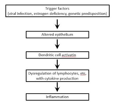
Sjögren syndrome   Updated: 02/13/2021 © Jun Wang, MD, PhD General features Third most common rheumatic disorder, after rheumatoid arthritis and systemic lupus erythematosus Typically presents with xerostomia , keratoconjunctivitis sicca (dry eye), rheumatoid arthritis, and hepergammaglobulinemia More common in women , average onset between 40-60 May involve lymph nodes, lung, kidney, etc Associated with Graves disease , Hashimoto thyroiditis , non-Hodgkin lymphoma, especially MALT lymphoma Pathogenesis Clinical features Xerostomia ( dry mouth ) Keratoconjunctivitis sicca ( dry eyes ) Enlarged parotid glands Rheumatoid arthritis Hypergammaglobulinemia Diagnostic criteria Inclusion criteria: At least one symptom of ocular or oral dryness Exclusion criteria History of head and neck radiation treatment Active hepatitis C infection Acquired immunodeficiency syndrome Sarcoidosis Amyloidosis Graft-verses-host disease IgG4-related disease Pathological

Pleomorphic adenoma

Pleomorphic adenoma   Updated: 02/13/2021 © Jun Wang, MD, PhD General features AKA Benign mixed tumor Most common tumor of salivary glands Most common in parotid gland More common in women Risk factors for malignant transformation: submandibular location, older age, larger size, radiation exposure Pathogenesis Unclear etiology Prior radiation increases risk Probably associated with Simian virus (SV40) Clinical features Painless, slow growing tumor Usually NOT involving facial nerve Rapid growth if malignant transformation ( carcinoma ex pleomorphic adenoma ) Pathological features Single firm, mobile, well-circumscribed mass Triphasic population of benign ductal cells, myoepithelial cells and stromal cells Fibrous capsule Ductal and myoepithelial components : Commonly glandular , may be squamous, spindled or oval, myoepithelial cells as the outer layer Mesenchymal components : Various, may be myxoid, hyaline, chondroid , adipose or osseous; mucin

Carcinoma ex pleomorphic adenoma

Carcinoma ex pleomorphic adenoma   Updated: 02/13/2021 © Jun Wang, MD, PhD General features Develops in association with benign primary or recurrent pleomorphic adenoma More common in older population History of previous pleomorphic adenoma required for diagnosis, unless co-existing pleomorphic adenoma identified Clinical features Sudden increase in growth, pain or facial paralysis, facial tingling, trismus Likely recur, metastasis Pathological features Infiltrating tumor with hemorrhage or necrosis Malignant components in the background of pleomorphic adenoma Most common adenocarcinoma with irregular glands, atypical cells, necrosis, active mitosis, etc Management Surgery Radiotherapy, chemotherapy, possibley WT1 based immunotherapy, etc Prognosis depends on extent, staging, grade, etc Back to salivary glands pathology Back to contents

Adenoid cystic carcinoma

Adenoid cystic carcinoma   Updated: 02/13/2021 © Jun Wang, MD, PhD General features Most common in minor salivary glands More common in 5 th to 6 th decades, men May harbor MYB-NFIB rearrangement Clinical features Slow growing, but aggressive Pathological features Small, poorly circumscribed or encapsulated and infiltrative Biphasic tumor with ductal and myoepithelial cells Benign appearing basaloid cells forming cribriform , solid or tubular pattern Commonly with perineural invasion Management Surgery Radiation Chemotherapy Back to salivary glands pathology Back to contents

Acinic cell carcinoma

Acinic cell carcinoma   Updated: 02/13/2021 © Jun Wang, MD, PhD General features #2 childhood salivary gland malignancy after mucoepidermoid carcinoma May be multicentric or bilateral Likely recur if incompletely excised Clinical features Usually parotid and minor salivary glands, also parotid lymph nodes Pathological features Solid , microcystic , papillary cystic, follicular Tumor cells with basophilic cytoplasm and small round nuclei Express cytokeratin, alpha-1-antichymotrypsin , alpha amylase Management Surgery Radiation Back to salivary glands pathology Back to contents

Pathology of salivary glands

Pathology of salivary glands Updated: 02/20/19 © Jun Wang, MD, PhD Key anatomy and histological features Major salivary glands Parotid gland: Predominantly serous glands Submandibular gland: Mixed serous glands and mucinous glands Sublingual gland: Predominantly mucinous glands Minor salivary glands Xerostomia Sialadenitis Sjogren syndrome Mucocele Salivary gland neoplasms Pleomorphic adenoma Carcinoma ex pleomorphic adenoma Warthin tumor Mucoepidemoid carcinoma Adenoid cystic carcinoma Acinic cell carcinoma Practice questions Back to contents