Posts

Medullary cystic kidney disease

Medullary cystic kidney disease   Updated: 10/04/2018 © Jun Wang, MD, PhD General features Autosomal dominant AKA autosomal dominant interstitial kidney disease (ADIKD) or autosomal dominant tubulointerstitial kidney disease (ADTKD) No extrarenal involvement Three subtypes Uromodulin kidney disease (UKD) ADTKD DUE TO MUTATIONS IN THE REN GENE (ADTKD-REN) Mucin-1 kidney disease (MKD) Pathogenesis Loop of Henle deficiency due to uromodulin mutation (UKD) Accumulation of prerenin in tubules due to renin mutation (ADTKD-REN) Abnormal intracellular localization of mucin-1 in renal tubules (MKD) Clinical features Family history of kidney disease and gout Adult-onset progressive renal failure Early onset gout (teenage years) Pathological findings Similar to nephronophthisis Back to kidney masses Back to contents
Nephronophthisis

Image
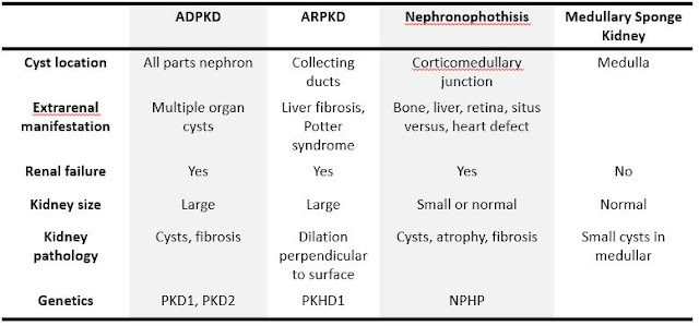
Nephronophthisis   Updated: 09/30/2021 © Jun Wang, MD, PhD General features Heterogeneous group Autosomal recessive Most common genetic cause of pediatric end stage kidney disease Characterized by corticomedullary cysts, atrophy and interstitial fibrosis Diagnosis based on clinical findings and confirmed by genetic testing Key pathogenesis Cilia/basal body abnormality Abnormal renal tubular development and function Clinical presentations Infantile Most severe form Bilateral disease Median age for renal failure: 1 year of age Extrarenal presentations common: retinitis pigmentosa, hepatic fibrosis, skeletal / CNS malformations, situs inversus, etc Juvenile form Most common Polyuria/polydipsia due to cortical and tubulointerstitial damage Progresses to chronic renal failure in 5-10 years Anemia and growth retardation May be associated with retinitis pigmentosa Adolescent form : Similar to juvenile with later presentation (median age renal failur

Multicystic Renal Dysplasia

Multicystic Renal Dysplasia   Updated: 10/05/2020 © Jun Wang, MD, PhD General features AKA multicystic dysplastic kidney Congenital Most common pediatric cystic renal disease Usually sporadic but may be familial Bilateral involvement usually results in stillbirth or death within the first few days of life  Pathogenesis Abnormal induction of metanephric mesenchyme by ureteral bud Clinical features No significant clinical problem if unilateral Abdominal mass: Most common cause of abdominal mass in the newborns Commonly associated with ureteropelvic obstruction, ureteral agenesis, atresia or reflux May presents with urinary tract infection, voiding dysfunction, or hypertension Radiologic findings Sonographic findings: Multiple cysts Voiding cystourethrography: Vesicoureteral reflux Dimercaptosuccinic acid (DMSA) renal scanning: Loss of renal function, used if sonographic examination inconclusive Pathological findings Commonly unilateral with con

MiT translocation subgroup carcinomas

MiT translocation subgroup carcinomas   Updated: 10/07/2020 © Jun Wang, MD, PhD General features MiT: microphthalmia associated transcription factors Gene fusion involving TFE3 transcription factor on Xp11.2 or TFEB gene fusion due to t(6;11) Diagnosis requires FISH or PCR More common in women More common in pediatric and young adult population Aggressive clinical course Pathological features TFE3 rearrangement Papillary and nested growth pattern Eosinophilic and clear discohesive pseudostratified cells t(6:11) TFEB  Biphasic, small and large tumor cells Markers Positive: TFE3, TFEB May be positive: HMB45, melan A Genetic abnormality Translocation involving X11p: TFE3 transcription facto t(6:11): TFEB Diagnosis FISH or PCR Back to renal cell carcinoma Back to kidney masses Back to contents

Medullary Sponge Kidney

Image
Medullary Sponge Kidney   Updated: 09/30/2021 © Jun Wang, MD, PhD General features Sporadic Rarely progress to end stage renal disease due to complications (infections, nephrolithiasis) May be associated with hemihypertrophy of body, Marfan’s, Caroli’s and Ehlers-Danlos syndrome Etiology and pathophysiology Likely developmental abnormality Clinical features Usually asymptomatic with normal renal function Commonly incidental finding by image studies Adult onset of calcifications, stones, hematuria and infection May have hypercalcemia Pathological findings Small cystic dilation of medullary collecting ducts Normal cortex Diagnosis Intravenous pyelography or CT: R enal tubular ectasia Differential diagnosis Management Treatment of stones Treatment of flank pain Monitor renal function Back to kidney masses Back to contents

Hypoplasia of kidney

Hypoplasia of kidney   Updated: 10/05/2020 © Jun Wang, MD, PhD General features Congenital smaller kidney Unilateral or bilateral Reduced number of nephrons Hypertrophied nephron Often with dysplasia (hypodysplasia) Key pathogenesis Probably developmental defect of metanephric renal blastema Clinical features Oligohydramnios Presentations of renal impairment May develop end-stage renal disease Key morphological features Reduced number of nephrons Normal architecture Hypertrophic glomeruli/tubules with Thickened Bowman’s capsule Abnormal glomerular basement membrane Dysplasia common Diagnosis Image studies Back to kidney masses Back to contents

Horseshoe kidney

Horseshoe kidney Updated: 10/03/2018 © Jun Wang, MD, PhD General features Most common congenital kidney anomaly Most common fused at lower poles If complete fusion: pancake kidney (formless mass in the pelvis) May have anomalous superior vena cava May develop cancers, renal cell CA most common, followed by urothelial CA and sarcoma Risks for Wilms tumor and carcinoid Key pathogenesis Unclear Mechanical fusion: Touch and fusion Teratogenic: Abnormal migration of nephrogenic cells Clinical features Commonly asymptomatic Hydronephrosis common Symptoms associated with obstruction, stones, infection Urinary tract infection: Most common symptom in children Diagnosis Sonography Voiding cystourethrogram for vesicoureteral reflux Evaluations Renal function Back to kidney masses Back to contents