Practice question answers papulosquamous disorders
Practice question answers
Papulosquamous disorders
Updated: 03/05/2019
© Jun Wang, MD, PhD
1. C. Well-demarcated plaque with erythematous base and
silver scales is most likely psoriasis,
as confirmed by epidermal hyperplasia with elongated rete ridges and absence of
granular layers. Dermatophytosis
usually has central resolution, peripheral scaling and annular shape, with
microscopic findings of neutrophilic infiltration and fungal hyphae identified
by special stains. Lichen
planus usually presents with pruritic, polygonal purple papules with
flat top, and microscopically it has lichenoid inflammation. Seborrheic
dermatitis usually presents as erythematous papules and plaques, with
yellowish scales, involving seborrheic areas, such as face, especially nasolabial
areas. Microscopically it has spongiosis centered on follicles, but usually not
elongated rete ridges. Squamous cell carcinoma has cytological atypia.
2. A. Psoriasis
is caused by cornification defects, either due to elevated cytokines, or LCE3
deletion. Autoimmune destruction of keratinocytes can be seen in various
dermatosis, including lichen planus,
erythema
multiforme and allergic
contact dermatitis. Chronic inflammation due to fungal infection is
seen in dermatophytosis.
Malignant transformation caused by UV light is the major cause of skin cancers.
Treponema pallidum infection is associated with syphilis.
3. C. Psoriasis
is caused by cornification defects, either due to elevated cytokines, or LCE3
deletion. BRAF mutation can be seen in various tumors, including melanocytic
nevus and melanoma. FGFR3 abnormality is associated with urothelial
papilloma, and certain type of urothelial
carcinoma. P53 mutation can be seen in many different cancers, especially
high grade cancers. PTCH mutation is seen in basal cell carcinoma.
4. A. Droplike erythematous to salmon-pink papules
with fine scales and microscopic findings of epidermal hyperplasia and Munro
microabscesses is most likely guttate psoriasis.
Lichen planus
usually presents with pruritic, polygonal purple papules with flat top, and
microscopically it has lichenoid inflammation. Pityriasis
rosea has herald patch followed by Christmas tree pattern of erythematous
patches with fine pink scale, and microscopic findings of spongiosis, but NOT
prominent epidermal hyperplasia. Seborrheic
dermatitis usually presents as erythematous papules and plaques, with
yellowish scales, involving seborrheic areas, such as face, especially nasolabial
areas. Microscopically it has spongiosis centered on follicles, but usually not
elongated rete ridges. Secondary syphilis
has prominent plasma cell infiltrates.
5. D. Widespread pustules and epidermal hyperplasia
with parakeratosis and numerous neutrophilic aggregates, especially at the
thinned granular layer, is likely to be pustular psoriasis,
especially when the patient has nail changes that are commonly seen associated with
psoriasis.
Dermatitis
herpetiformis has clusters
of small pruritic vesicles or pustules, and microscopically it has subepidermal
vesicle with neutrophilic aggregates. Pityriasis
rosea has herald patch followed by Christmas tree pattern of erythematous
patches with fine pink scale, and microscopic findings of spongiosis, but NOT
prominent epidermal hyperplasia. Secondary syphilis
has prominent plasma cell infiltrates.
6. B. Psoriasis
is caused by cornification defects, either due to elevated cytokines, or LCE3
deletion. Autoimmune destruction of keratinocytes can be seen in various
dermatosis, including lichen planus,
erythema
multiforme and allergic
contact dermatitis. Chronic inflammation due to fungal infection is
seen in dermatophytosis.
Hyperglycemia as seen in diabetes
is associated with increased risk of infections, but not psoriasis.
Patient with malnutrition has symptoms and signs such as weight loss, anemia,
etc.
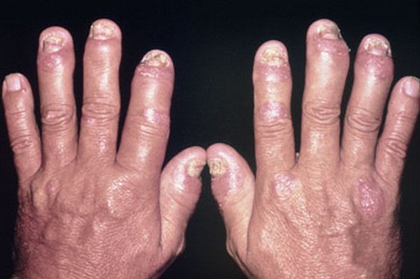
7. B. Small joint tenderness and swelling in a patient
with psoriasis,
is most likely psoriatic arthritis (sausage
fingers). Infectious arthritis usually has leukocytosis. Rheumatic and
rheumatoid arthritis have abnormal serology studies.
8. C. Conjunctivitis in a patient with psoriasis,
is most likely psoriatic conjunctivitis. The diagnosis of psoriasis
is confirmed by biopsy of the skin lesion.
9. D. Seborrheic
dermatitis usually presents as erythematous papules and plaques, with
yellowish scales, involving seborrheic areas, such as face, especially nasolabial
areas. Atopic
dermatitis has diffuse facial scaly rash with erythematous base that
spares nasal area. Pemphigus
erythematosus and SLE have malar rash on face. Psoriasis
has well-demarcated papule/plaque with erythematous base and silver scales.
10. D. Seborrheic
dermatitis is associated with abnormal immune activity to normal
fungal colonization. Autoimmune process is associated with various dermatosis,
including pemphigus
and bullous
pemphigoid. Cornification defects are seen in psoriasis.
Viral infection can be associated with various dermatosis, including herpes,
pityriasis
rosea and erythema
multiforme.
11. B. Lichen
planus usually presents with pruritic, polygonal purple papules with
flat top, and microscopically it has lichenoid inflammation. Dermatophytosis
usually has central resolution, peripheral scaling and annular shape, with
microscopically identified fungal hyphae. Pityriasis
rosea has herald patch followed by Christmas tree pattern of erythematous
patches with fine pink scale, and microscopic findings of spongiosis, but NOT lichenoid
inflammation. Pustular
psoriasis has widespread pustules and epidermal hyperplasia with
parakeratosis and numerous neutrophilic aggregates, especially at the thinned
granular layer. Secondary syphilis
has prominent plasma cell infiltrates.
12. A. Clumped IgM deposit at dermoepidermal junction
is seen in lichen
planus. IgG deposit at intercellular junctions of keratinocytes,
where desmosomes are, is seen in pemphigus.
Granular deposit of IgA at dermal papillae is seen in dermatitis
herpetiformis. Linear deposit of IgG at dermoepidermal junction is
seen in bullous
pemphigoid.
13. B. Koebner
phenomenon is defined as new skin lesion on areas of cutaneous injury. The etiology is unknown.
14. C. It is not uncommon for lichen
planus to be associated with hepatitis C. There is recommendation to
test for hepatitis C in patients with lichen
planus.
15. B. Lichen
planopilaris is lichenoid inflammation involving hair follicles. It is
commonly associated with alopecia. Dermatophytosis
usually has central resolution, peripheral scaling and annular shape, with
microscopically identified fungal hyphae. Psoriasis
has well-demarcated papule/plaque with erythematous base and silver scales, and
microscopically, epidermal hyperplasia with elongated rete ridges and absence
of granular layers. Seborrheic
dermatitis usually presents as erythematous papules and plaques, with
yellowish scales, involving seborrheic areas, such as face, especially nasolabial
areas. Microscopically it has spongiosis centered on follicles, but usually not
elongated rete ridges. Secondary syphilis
has prominent plasma cell infiltrates.
16. A. Lichen
planus is CD8+ T cell-mediated immune reaction against basal
epidermal cells or dermoepidermal junction, with unknown triggering factor. Psoriasis
is caused by cornification defects, either due to elevated cytokines, or LCE3
deletion. Seborrheic
dermatitis is associated with abnormal immune activity to normal
fungal colonization. Viral infection can be associated with various dermatosis,
including herpes,
pityriasis
rosea and erythema
multiforme.
17. C. See discussion of question 4.
18. B. Pityriasis
rosea is usually associated with certain viral infection, including vaccines.
Certain human herpes virus, such as HHV6 and HHV7, may cause pityriasis
rosea. But there is no known association between pityriasis
rosea and human herpes virus 1. Malassezia is associated with seborrheic
dermatitis. Treponema pallidum is associated with syphilis.
There is no known associated between pityriasis
rosea and either EBV or HPV.
19. E. Non-pruritic skin rashes in a patient with history
of glans ulcer is highly suspicious for secondary syphilis,
which is further supported by diffuse dermal plasma cell infiltrate in this
case. Also see discussion of question 4.
20. E. The screening test for syphilis
include Rapid plasma regain (RPR) and Venereal Disease Research Laboratory
(VDRL), and the confirmation tests include Fluorescent treponemal
antibody-absorption (FTA-ABS) or Treponema pallidum hemagglutination assay. Culture
is for bacterial infections. Flow cytometry studies is more useful for
hematopoietic disorders, especially neoplasia. Monospot is for infectious
mononucleosis. RT-PCR for HIV RNA is for HIV infection, which is a high
risk for this patient, but irrelevant to his skin rash.
21. A. This is a silver that highlighted fungal
hyphae. Dermatophytosis
may have various microscopic pattern but fungal hyphae identified by special
stains is confirmative for the diagnosis.
Back to papulosquamous
disorders
Back to contents
Comments
Post a Comment