Posts

Showing posts with the label uterus

Malignant mixed müllerian tumor

Malignant mixed müllerian tumor   Updated: 12/16/2020 © Jun Wang, MD, PhD General features Biphasic tumor with malignant epithelial and stromal components (carcinosarcoma) Postmenopausal women Associated with chronic estrogen stimulation Highly aggressive Clinical presentations Asymptomatic  Or triad of pain, uterine bleeding, and rapid enlargement of uterus Pathological findings Epithelial components: Atypical cells with glandular differentiation Stromal components: Atypical poorly differentiated spindle cells with high nuclear/cytoplasmic ratio and hyperchromic irregular nuclei Necrosis and mitosis Laboratory finding Elevated Serum CA125 Treatment Surgery Chemotherapy Radiation therapy Back to female genital tract Back to contents
Endometritis

Endometritis   Updated: 12/16/2020 © Jun Wang, MD, PhD General features Inflammation of endometrium Pregnancy-related or unrelated to pregnancy Acute and chronic May be associated with salpingitis, oophoritis and peritonitis Clinical presentations Acute endometritis : symptoms of infection, fever, septic shock , etc Chronic endometritis : usually asymptomatic Etiology Acute endometritis : bacterial infection, retained tissue or instrumentation Chronic endometritis : Pelvic inflammatory disease, IUD, postpartum, post-abortion (retained tissue), infection, etc Key pathological findings Acute endometritis : neutrophilic infiltration, microabscess Chronic endometritis : Plasma cells in stroma Diagnosis Usually based on clinical findings Treatment Acute endometritis: Antibiotics, removing retained tissue Chronic endometritis: Antibiotics, removing retained tissue, treating underlying causes Back to female genital tract Back to contents

Endometriosis and adenomyosis

Endometriosis and adenomyosis   Updated: 12/16/2020 © Jun Wang, MD, PhD General features Benign endometrial mucosa (glands and stroma) in locations outside uterine cavity Chronic inflammatory, estrogen-dependent disease Reproductive age with active hypothalamic-pituitary-ovarian axis function Most common site: ovaries Adenomyosis : benign endometrial tissue in myometrium Associated with endometrioid type and clear cell type ovarian adenocarcinoma Clinical presentations Pain (dysmenorrhea) Infertility Key pathogenesis 4 hypothesis Regurgitation : Expulsion of endometrial tissue along fallopian tube during menstruation Benign metastasis : Spread of benign endometrial tissue in a similar means of tumor metastasis Extrauterine stem/progenitor cells : Extrauterine stem/progenitor cell differentiation in to endometrial tissue Metaplasia : Transformation of other type of tissue into endometrial tissue Key pathological findings Hemorrhagic nodules, chocolate

Serous carcinoma

Image
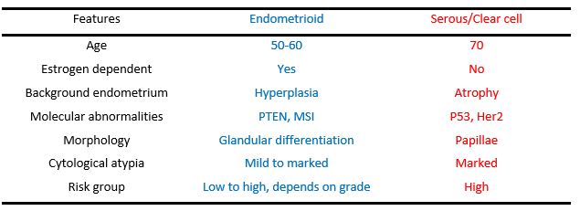
Serous carcinoma   Updated: 12/16/2020 © Jun Wang, MD, PhD General features Most common type 2 (estrogen independent) endometrial carcinoma In background of endometrial atrophy More common in black NOT associated with estrogen replacement therapy Associated with endometrioid endometrial adenocarcinoma , clear cell carcinoma and ovarian serous carcinoma Highly aggressive Pathogenesis Less defined Clinical presentations Asymptomatic or uterine bleeding Pathological findings Papillae Marked cytological atypia Necrosis Calcification Molecular abnormality p53 Her2 E-cadherin Comparison with type 1 endometrial adenocarcinoma Treatment Surgery Chemotherapy Radiation therapy Back to Adenocarcinoma of endometrium Back to female genital tract Back to contents

Endometrioid type adenocarcinoma

Image
Endometrioid type adenocarcinoma   Updated: 12/21/2020 © Jun Wang, MD, PhD General features Type 1 (estrogen dependent) endometrial carcinoma In background of endometrial hyperplasia Associated with estrogen replacement therapy More common in white Pathogenesis Prolonged estrogenic stimulation with reduced progestational activity Clinical presentations Asymptomatic or uterine bleeding Pathological findings Crowd irregular glands , possible fusion of glands Atypical cells Squamous metaplasia: benign squamoid changes, NOT counted as solid area for FIGO Weak p16 reactivity (strong p16 reactivity in serous carcinoma and endocervical adenocarcinoma ) FIGO grade I: well differentiated, <5% nonsquamous solid component II: moderately differentiated, 6-50% nonsquamous solid component III: poorly differentiated, > 50% nonsquamous solid component Upgrade with marked cytological atypia Molecular abnormality Loss of function of PTEN MSI: microsate

Endometrial stromal tumor

Endometrial stromal tumor   Updated: 12/16/2020 © Jun Wang, MD, PhD General features Benign: endometrial stromal nodule 5 th to 6 th decades Excellent prognosis Malignant: endometrial stromal sarcoma 40-50 years Rearrangements of JAZF1, SUZ12, PHF1, and EPC1, causing abnormal oncogene expression Commonly recur and metastasize Clinical presentations Benign: endometrial stromal nodule Asymptomatic, usually incidental finding Malignant: endometrial stromal sarcoma Abnormal uterine bleeding, pelvic pain/pressure, and/or uterine mass Pathological findings Benign: endometrial stromal nodule Well-circumscribed growth, may be polypoid Monotonous round endometrial stromal cells No cytological atypia Malignant: endometrial stromal sarcoma Infiltrating pattern Mild to severe cytological atypia Low-grade, high-grade, undifferentiated Necrosis Treatment Malignant: Surgery Chemotherapy Radiation therapy Back to female geni

Endometrial polyp

Endometrial polyp   Updated: 12/16/2020 © Jun Wang, MD, PhD General features Balanced growth of both glands and stroma Either hyperplastic or functional/secretory May be associated with tamoxifen therapy for breast cancer Clinical presentations Asymptomatic or uterine bleeding Key pathological findings Polypoid growth attached to endometrium Dilated glands, fibrous stroma, thick walledvessels No complex architecture No cytological atypia Back to female genital tract Back to contents

Endometrial hyperplasia

Endometrial hyperplasia   Updated: 12/16/2020 © Jun Wang, MD, PhD General features Over proliferation of endometrial glands Associated with polycystic ovarian disease (Stein-Leventhal syndrome), ovarian granulosa cell tumors (functional), ovarian cortical stromal hyperplasia, estrogen replacement therapy without progestational agents and high body mass index Classified according to architecture (simple vs. complex) and cytological features (with or without atypia) Simple hyperplasia : Usually no cytological atypia, slightly increased risk for endometrial carcinoma Complex hyperplasia : Commonly with cytological atypia , high risk of adenocarcinoma of endometrium Endometrial intraepithelial neoplasm A preferred term for atypical endometrial hyperplasia, precancerous lesion Diagnostic criteria Area of glands greater than stroma (volume percentage stroma less than 55%) Cytology differs between architecturally crowded focus and background Maximum linear dimensio

Dysfunctional uterine bleeding

Dysfunctional uterine bleeding   Updated: 12/22/2021 © Jun Wang, MD, PhD General features Definition: Bleeding >5 days of unknown cause in women of childbearing age Clinical, NOT pathology term Ovulate or anovulatory  Anovulatory most common Commonly due to endocrine disorders, such as polycystic ovary disease , ovarian failure, metabolic disorders, etc Higher risk for endometrial hyperplasia and carcinoma due to unbalanced estrogen effect Key pathogenesis Abnormal hormonal function Ovulatory: inadequate proliferative phase, inadequate secretory phase, irregular shedding or membranous dysmenorrhea Anovulatory: unbalanced estrogen effects, proliferative endometrium during chronological secretory phase, risk of adenocarcinoma of endometrium Key pathological findings Uneven differentiation of glands and stroma in a cycle, for example, mixed proliferative and secretory phase glands in a proliferative phase stroma Treatment Underlying causes Rule out othe

Clear cell carcinoma

Clear cell carcinoma   Updated: 12/16/2020 © Jun Wang, MD, PhD General features A type 2 (estrogen independent) endometrial carcinoma Post-menopausal Highly aggressive Pathogenesis Less defined Clinical presentations Asymptomatic or uterine bleeding Pathological findings Clear or hobnail cells Marked cytological atypia Molecular abnormality p53 Her2 E-cadherin Treatment Surgery Chemotherapy Radiation therapy Back to female genital tract Back to contents

Endometrial adenocarcinoma

Image
Endometrial adenocarcinoma   Updated: 11/03/2023 © Jun Wang, MD, PhD General features Most common gynecologic malignancy in US Incidence increasing More common in postmenopausal women Associated with complex endometrial hyperplasia, diabetes, dysfunctional uterine bleeding, hypertension, infertility, Muir-Torre syndrome, obesity, prolonged estrogen use, tamoxifen use Endometrioid type most common, followed by serous type May be part of Lynch Syndrome Pathogenesis Prolonged estrogenic stimulation with reduced progestational activity if endometrioid Clinical presentations Uterine bleeding Classification Type 1: Endometrioid endometrial adenocarcinoma Type 2: Serous carcinoma , Clear cell carcinoma Molecular changes PTEN: type 1, Endometrioid endometrial adenocarcinoma P53: type 2, Serous carcinoma , Clear cell carcinoma Risk groups Defined by stage, pathological features, etc Low-risk : Grade 1 endometrial cancer of endometrioid histology that i

Female genital tract

Pathology of female genital tract   Updated: 01/23/2024 © Jun Wang, MD, PhD Key anatomic features Opened channel, pathological basis for pelvic inflammatory disease Cervix: exocervix, endocervix, transformation zone Uterus: fundus, body Fallopian tube: intramural, isthmus, ampulla, infundibulum, fimbriae Placenta: umbilical cord, fetal membrane, placenta Key histologic features Stratified squamous epithelium from vulva to exocervix Simple columnar epithelium from endocervix to fimbriae Uterine corpus Endometrium: Glands, stroma Myometrium Perimetrium: Mesothelium/peritoneum Ovaries: three components Epithelium (mesothelium) Stroma Germ cells (follicles) Placenta: Cytotrophoblast, syncytiotrophoblast, intermediate trophoblast Infectious/inflammatory diseases Diseases of vagina and vulva Molluscum contagiosum Lichen sclerosus Paget disease Sarcoma botryoides Squamous cell carcinoma Diseases of cervix Cervical intraepithelial neopla

Lynch syndrome

Lynch syndrome   Updated: 11/03/2023 © Jun Wang, MD, PhD General features Hereditary non-polyposis colorectal cancer Autosomal dominant Most common hereditary colorectal carcinoma syndrome 80% of patients develop colorectal carcinoma Increased risk of cancers of endometrium , ovaries, small bowel, stomach , upper urinary tract , and brain, or skin ( Muir-Torre syndrome ) Genetic abnormalities Mismatch repair genes: MLH1, MSH2, MSH6, and PMS2 genes EPCAM: causing methylation and silencing of MSH2 MMRs MutSalpha: Heterodimer of MSH2 and MSH6, rocognizes base-pair mismatches MutLalpha: Heterodimer of MLH1 and PMS2, promote excision of mismatches Diagnostic approach Screening test recommendations  CRC diagnosed at age <50 years Synchronous CRC, metachronous CRC, or Lynch syndrome-associated tumors CRC with the MSI-H histology diagnosed at age <60 years CRC in ≥1 first-degree relative with a Lynch syndrome-related tumor with 1 of the cancers diagnosed at ag

Leiomyosarcoma

Leiomyosarcoma   Updated: 08/07/2020 © Jun Wang, MD, PhD General features Malignant tumor of smooth muscle Commonly in uterus, skin, subcutis, deep soft tissue and GI Third most common retroperitoneal sarcoma after liposarcoma and undifferentiated pleomorphic sarcoma Aggressive clinical behavior Most common metastasis site: lung Clinical presentations Mass Symptoms depending on location Key morphological features Large tumor Hypercellular, hyperchromic spindle cells Pleomorphic cells, usually marked cytological atypia Necrosis, active mitosis, atypical mitosis Markers Positive for desmin, smooth muscle actin Genetic abnormalities Complex, commonly p53 mutation and p16 overexpression Treatment Excision Chemotherapy Radiation Poor prognosis indicators Retroperitoneal, mesenteric or other deep location > 5 cm, except uterus > 65 years C-myc expression Back to soft tissue tumors Back to contents

Leiomyoma

Leiomyoma   Updated: 08/07/2020 © Jun Wang, MD, PhD General features Benign tumor of smooth muscle Commonly in uterus, skin, subcutis, deep soft tissue and GI If multiple: Leiomyomata Clinical presentations Mass Uterine leiomyoma may cause dysfunctional uterine bleeding or lower abdomen pressure-related symptoms Key morphological features Well circumscribed firm mass Bulging trabecular cut surfaces Bundles or fascicles of spindled cells with minimal atypia Cigar shaped nuclei Usually no necrosis, no mitosis Markers Positive for desmin, smooth muscle actin Genetic abnormalities HMGIC, HMGIY, MED12 for uterine leiomyoma Treatment Excision Back to soft tissue tumors Back to contents