Terms of skin disorders
Terms
of skin disorders
Changes of darkness of skin:
Flat colored lesions:
Purulent: Containing or discharging pus
Palisade: Monolayer of relatively long cells or organisms arranged loosely perpendicular to a surface and parallel to each other
Updated: 02/11/2022
© Jun Wang, MD, PhD
Clinical
Changes of darkness of skin:
- Hyperpigmentation: Darker than normal surrounding skin
- Hypopigmentation: Lighter than normal surrounding skin
Cyst: Encapsulated cavity or sac lined
by true epithelium
Flat colored lesions:
- Macule:
Circumscribed flat area of discoloration < 1 cm
- Patch: Flat area of discoloration > 1 cm
- Fissure: Crack through the epidermis and into the dermis, NO tissue loss
- Erosion: Discontinuity of skin causing partial loss of epidermis
- Excoriation: Deep linear scratch, commonly self-induced
- Ulceration: Discontinuity of skin causing complete loss of epidermis and possible loss of dermis
Lichenification: Thickening of skin due to chronic
irritation (rubbing), may have discoloration
Onycholysis: Separation of nail plate
Poikiloderma: Skin atrophy with hypopigmentation and telangiectasia
Raised
lesions:
- Papule: Elevated flat topped area, < 10 mm
- Plaque: Elevated flat topped area, > 10 mm
- Nodule: Solid, palpable lesion > 10 mm
- Tumor: Solid palpable lesion > 2 cm
Red
discoloration:
- Erythema: Redness of skin due to increased blood flow, NO extravasation of blood cells, may blanch
- Purpura: Red discoloration of skin or mucosa, caused by extravasation of red blood cells
Scale: Dry, plate-like excrescence,
associated with excess stratum corneum
Telangiectasia: Visible persistent dilation of
small, superficial cutaneous blood vessels, may blanch
Vesicular
lesions:
- Vesicle: Fluid filed area, < 10 mm
- Bullae: Fluid filled area > 10 mm
- Blister: Vesicle or bullae
- Pustule: Blister filled with pus
Wheals: Itchy, transient, elevated area
with variable blanching and erythema, due to dermal edema
Special clinical terms
Histology
Ash-leaf spots: hypopigmented
macules, which are usually elliptic in shape, seen in tuberous sclerosis
Shagreen
patches: Orange-peel–textured area of connective tissue hamartoma most commonly
over the lower trunk
Histology
Abnormal
intercellular connections:
- Acantholysis: Loss of intercellular connections (desmosomes) between keratinocytes
- Spongiosis and vesicles: Widened intercellular or dermo-epidermal junctions
Abnormal
keratosis:
- Dyskeratosis: Premature kerainization of keratinocytes below granular layer, often with eosinophilic cytoplasm
- Hyperkeratosis: Thickened cornified layer, often with prominent granular layer
- Hypergranulosis: Thickened granular layer, associated with intensive irritation (rubbing)
- Parakeratosis: Retaining nuclei of cells in cornified layer, nonspecific, associated with chronic irritation
Acanthosis: Thickening of epidermis
Atypia: Abnormal structural architecture
or cellular morphology, including shape, size, and staining pattern, either reactive or neoplastic
Basaloid: With the appearance of basal layer
epidermis, usually less cytoplasm, hyperchromatic nuclei
Cyst: Material-containing space lined by
epithelium
Elastosis: Grayish discoloration of dermis,
usually due to sun damage, commonly seen in sun-damaged skin, associated with
actinic keratosis, and other skin cancers
Epithelioid: With the appearance of epithelial
cells, usually round to oval nuclei
Hyperchromasia:
Darker than normal nuclear stain, usually due to increased quantity of DNA, seen in dysplasia
Intercellular bridge: Thread-like connection between widened intercellular spaces, containing
desmosome, characteristic for squamous differentiation
Koilocyte: Changes of squamous cells characterized by enlarged nuclei with various degree of hyperchromasia, irregular nuclear membrane contour, and perinuclear halo (a clear area around nuclei), commonly associated with human papillomavirus infections
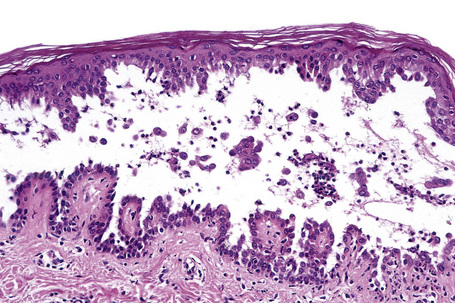
Pagetoid spread: Spread of tumor cells with relatively paler
cytoplasm, either singly or in cluster, among benign epidermal keratinocytes
with relatively darker cytoplasm, as seen in Paget disease.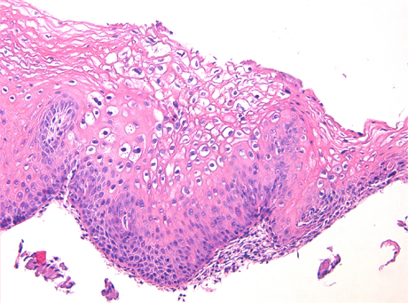
Palisade: Monolayer of relatively long cells or organisms arranged loosely perpendicular to a surface and parallel to each other
Pleomorphism:
Variations of morphology, usually a sign of high-grade tumor cells
Psoriasiform
hyperplasia: Regular elongation of rete ridges and suprapapillary thinning,
nonspecific, seen at psoriasis, or results of chronic irritation
Squamous pearl: Keratinized structure with concentric layers of atypical squamous cells, usually seen in squamous cell carcinoma
Squamous pearl: Keratinized structure with concentric layers of atypical squamous cells, usually seen in squamous cell carcinoma
Back
to contents
Comments
Post a Comment