Posts

Showing posts from July, 2018

Ganglion cyst

Ganglion cyst Updated: 07/31/2018 © Jun Wang, MD, PhD General features Small cyst-like lesion near joints, tendons Very common Clinical presentations Likely asymptomatic Pain or swelling Limited activity Pathogenesis Mucoid degeneration of joint capsule, tendon or tendon sheath Pathological features Mucus filled cystic lesion  No epithelial lining Treatment Surgery Back to bone tumors Back to contents

Fibrous dysplasia

Fibrous dysplasia Updated: 07/31/2018 © Jun Wang, MD, PhD General features Developmental, non-neoplastic disorder of bone-forming mesenchyme Usually begins prior to puberty and grows slowly Slightly more common in men Malignant transformation: Rare, may represent dedifferentiation of low-grade osteosarcoma; may occur after radiation therapy Clinical presentations Deformities May have pain or swelling Pathogenesis Bone maturation arrest at the woven bone stage Most common sites Medulla of diaphysis or metaphysis of craniofacial bones, femur, ribs Key radiologic findings Lytic lesion in the metaphysis or diaphysis  "Ground glass" appearance Key morphological features Irregular trabeculae of metaplastic woven bone (never matures) Hypocellular, fibroblastic stroma Genetic abnormality Probably activating point mutation (possibly a submit of GNAS1), resulting elevated intracellular cyclic adenosine monophosphates (cAMP) Treatment

Fibrous cortical defect/nonossifying fibroma

Fibrous cortical defect/nonossifying fibroma Updated: 07/01/2023 © Jun Wang, MD, PhD General features AKA metaphyseal fibrous defect Often less than 1 cm Called nonossifying fibroma if loose, > 5 cm and associated with intramedullary component Affects teenagers Usually resolves in a few years, replaced by cortical bone Likely to be multiple Clinical presentations Usually incidental finding Asymptomatic, possibly pain Pathogenesis Unclear, probably developmental defect Most common sites Metaphysis of distal femur, tibia Key radiologic findings Sharply demarcated radiolucency with zone of sclerosis NO periosteal reaction Key morphological features Loose storiform pattern spindle cell proliferation Scattered giant cells Treatment Surgery Back to bone tumors Back to contents

Tenosynovial giant cell tumor

Tenosynovial giant cell tumor Updated: 07/31/2018 © Jun Wang, MD, PhD General features Benign Diffuse (pigmented villonodular synovitis) or localized (giant cell tumor of tendon sheath) Usually monoarticular Locally aggressive Clinical presentations Usually painless mass Partial disability of joint possible Pathological features Numerous multinuclear giant cells Proliferating histiocytes Treatment Surgery Back to bone tumors Back to contents

Ewing sarcoma

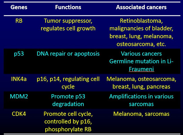
Ewing sarcoma Updated: 08/12/2022 © Jun Wang, MD, PhD General features AKA primitive neuroectodermal tumor (PNET) #2 bone sarcoma in children, after osteosarcoma Usually whites, ages 5-20 years Possible multiple lesions at presentation 5 year survival near 75% Clinical presentations Pain, fever, weight loss Most common sites Medullary cavity, usually shaft Femur, tibia, humerus, etc Key pathogenesis EWS-Fli1 activates Cyclin D and Cyclin E Activation of Cyclin D and Cyclin E promotes cell cycle progression through suppression of tumor suppressor RB Key radiologic findings Mottled, osteolytic lesion with sunburst periosteal reaction Lamellated periosteal reaction Codman’s triangle : Shadow between cortex and raised ends of periosteum (due to reactive bone formation), non-specific (can be seen in osteosarcoma ) Key morphological features Sheets of small blue cells with scant cytoplasm and round nuclei Markers Positive for CD99 Differe

Aneurysmal bone cyst

Aneurysmal bone cyst Updated: 07/31/2018 © Jun Wang, MD, PhD General features Uncommon Common ages 1-20 years, no gender preference May be secondary to trauma or other bone lesions Benign but grows rapidly May recur after curettage Clinical presentations Localized pain and swelling Pathological fracture Most common site Metaphysis of long bones Posterior vertebrae Key radiologic findings Expansile, lytic metaphyseal  "Eggshell" sclerotic rim Key morphological features Large cystic spaces separated by septa containing giant cells and spindle cells Cysts may be filled with blood No endothelial lining cysts Genetic abnormality USP6 overexpression Treatment Surgery Back to bone tumors Back to contents

Giant cell tumor of bone

Giant cell tumor of bone Updated: 07/31/2018 © Jun Wang, MD, PhD General features Also called osteoclastoma Benign but locally aggressive More common: 20-40 years, women, oriental countries Associated with Paget disease of bone Usually good prognosis Clinical presentations Pain, swelling, limitation of joint movements Pathologic fracture Most common site Knees, usually epiphysis Pathogenesis RANKL expressed by tumor cells recruit circulating monocytes to form osteoclasts Key radiological findings Soap bubble appearance Key morphological features Expansile lytic lesion   Numerous multinucleated giant cells Background of uniform mononuclear cells Treatment Surgery Radiation if excision impossible Back to bone tumors Back to contents

Chondrosarcoma

Chondrosarcoma   Updated: 08/12/2022 © Jun Wang, MD, PhD General features Malignant tumors wiht complete cartilage matrix May arise from osteochondroma Third most common bone malignancy after myeloma and osteosarcoma Slightly more common in male, >40 Classification: Conventional (central, peripheral, juxtacortical/periosteal, most common type) and variants (clear cell, dedifferentiation, mesenchymal, myxoid) Dedifferentiated chondrosarcoma: Coexistence of well differentiated (low grade) cartilaginous component and high grade anaplastic component, high grade, poor prognosis Clinical presentations Deep dull achy pain Pain at night Most common sites Axial skeleton, especially pelvis and ribs Key radiological findings Destructive Expansile With or without ossification Key morphological features Destructive tumor with bluish-white cartilaginous tissue Permeating growth suggested by bony trabeculae entrapment Treatment Surgery Possibly therapies

Chondroma

Chondroma   Updated: 08/12/2022 © Jun Wang, MD, PhD General features Benign cartilaginous tumor Including enchondroma (diaphyseal medullar cavity), subperiosteal/juxtacortical chondroma and soft tissue chondroma Clinical presentations Asymptomatic Pain Pathological fracture Most common sites Small bones of hands and feet Femur, proximal humerus Usually not in flat bones, in contrast to chondrosarcoma Key radiological findings Lytic expansile lesion Key morphological features Well circumscribed cartilage growth Hyaline cartilage No invasion Genetic abnormalities HMGA2 / HMGI-C IDH1 and IDH2 in enchondromas Maffucci syndrome: Very rare Somatic IDH1 mutation, non hereditary Multiple enchondromas, hemangioma (subcutaneous), and soft tissue lymphangiomas Short stature, underdeveloped muscles and bone deformities Higher risk for ovarian and liver carcinomas, brain gliomas Ollier disease Enchondromas of long bone and flat bones IDH

Osteochondroma

Osteochondroma Updated: 09/05/2018 © Jun Wang, MD, PhD General features AKA exostosis Most common benign bone tumor More common in male, < 20 Growing stopped and lesion ossified at puberty May transform into chondrosarcoma Clinical presentations Slow growing Pain if affect nerves or stalk is broken Most common site Metaphysis Not medullary cavity Distal femur, proximal tibia Not in intramembranous bones Key radiological findings Pedunculated Often point away from the nearest joint Cortex and medulla continuous with underlying bone Key morphological features Cartilaginous cap with disorganized growth plate and ossification towards base Relative normal bone trabeculae and marrow Genetic abnormalities Seen in hereditary exostoses ( Hereditary multiple osteochondromas ) Various including EXT1 or EXT2 Treatment Simple excision Back to bone tumors Back to contents

Osteosarcoma

Image
Osteosarcoma Updated: 09/24/2018 © Jun Wang, MD, PhD General features Any malignant tumors that produce osteoid directly from tumor cells Most common primary bone tumor after myeloma Young age, 10-25 Most common metastasis site: Lung Rare metastasis to lymph nodes Risk factors If > 40 Paget disease Radiation exposure Thorotrast Childhood chemotherapy Other bone disorders such as fibrous dysplasia, osteochondromatosis, chondromatosis, etc Clinical presentations Most common clinical presentation: Localized pain Mass Most common site Metaphysis of long bones, especially around the knee Medullary cavity Key pathogenesis Unclear, probably associated with rapid bone growth Key laboratory findings Elevated alkaline phosphatase Key radiological findings Large, destructive mass Lytic or blastic Permeative margins Sunburst pattern due to bone formation Codman’s triangle : Shadow between cortex and raised ends of periosteum (due to react

Osteoblastoma

Image
Osteoblastoma Updated: 07/27/2018 © Jun Wang, MD, PhD General features Also called giant osteoid osteoma Usually > 2 cm Slightly more common in female, mean age 20 years old Most common sites: Spine and sacrum Clinical presentations Pain, worse at night, NOT relieved by NSAIDs Key pathogenesis Progress pain, NOT respond to aspirin Key radiological findings Radiolucent nidus Surrounding bony sclerosis and cortical thickening No invasion Key morphological features Anastomosing osteoid trabeculae and woven bone rimmed by osteoblasts No invasion No atypia Differential diagnosis Treatment Curettage En Bloc resection Back to bone tumors Back to contents

Osteoiod osteoma

Image
Osteoid osteoma Updated: 07/27/2018 © Jun Wang, MD, PhD General features Benign Usually < 1.5 cm More common in male, 5-24 years old Limited growth Most common sites: Long bones, especially femur and tibia Clinical presentations Pain, worse at night, relieved by NSAIDs Key pathogenesis Pain associated with prostaglandin E2 or nerves in reactive zone Key radiological findings Radiolucent nidus with surrounding bony sclerosis and cortical thickening No invasion Key morphological features Anastomosing osteoid trabeculae and woven bone rimmed by osteoblasts No invasion No atypia Differential diagnosis Treatment NSAIDs and other medical managements Excision Radiofrequency ablation Back to bone tumors Back to contents