Posts

Showing posts from January, 2024

Clostridium difficile

Clostridium difficile Updated: 02/03/2024 © Jun Wang, MD, PhD   General features Gram-positive, anaerobic, sporogenic bacterium May be part of normal colonic flora Most common cause of pseudomembranous colitis Most commonly nosocomial infectious diarrhea in the U.S. Colitis due to overgrowth and exotoxin production Results of interruption of normal colonic flora by antibiotics, chemotherapy or immunosuppression, etc Pathogenesis Disruption of normal colonic flora Colonization of C. Diff Production of exotoxins Toxin A: Enterotoxin, mucosal injury, fluid loss and inflammation, granulocyte attractant Toxin B: Cytotoxin, cytopathic Key clinical features Fever, abdominal pain and cramping Green foul-smelling diarrhea May perforate and cause septic shock Colonoscopic findings Colonic yellow plaques and nodules Pathologic findings Volcano or mushroom-like eruption of fibrin, mucin and inflammatory cells Diagnosis History of hospitalization and antibiotics

Vibrio Cholerae

Vibrio Cholerae Updated: 02/19/2024 © Jun Wang, MD, PhD   General features G-negative, facultative anaerobe and highly motile comma-shaped bacteria Lives in warm brackish water Copepods or shellfish in contaminated water Higher risk after natural disaster Human carriage may persist after untreated infection Key clinical features Rice-water stools with tremendous fluid loss ( Thin watery with flecks of mucus ) Hypovolemic shock if not treated Pathogenesis Attachment to intestinal mucosa Motility Mucinase Toxin co-regulated pili (TCP) Cholera enterotoxin (choleragen): similar to E. coli LT Activation of adenylate cyclase Increased cAMP Efflux of Cl – and H 2 O Diagnosis Presumptive diagnosis in patients with severe diarrhea Confirmation Isolation of v. cholerae from stool culture Grow on thiosulfate citrate bile sucrose agar or taurocholate tellurite gelatin agar Antigen detection Molecular testing Darkfield microscopy Management Adequate IV Fluid and

Shigella

Shigella Updated: 02/03/2024 © Jun Wang, MD, PhD   General features G-negative rods, non-motile, noncapsulated, facultatively anaerobic Highly virulent Leading cause of diarrheal death, with most common in south Asia and sub-Saharan Africa 4 groups: S. dysenteriae, S. flexneri, S. boydii, S. sonnei Key clinical features Fecal-oral Transmission Severity depends on age of patient and the strain S. dysenteriae type 1 with toxin most severe Fever (generally >101.0°F) Lower abdominal cramps; tenesmus; multiple scanty, bloody, mucoid stools Diarrhea: first watery, then bloody with WBCs Pathogenesis : Invasive but rarely causes septicemia Invade submucosa through M-cells (Microfold cells over lymphoid aggregates) Proliferate in macrophage Invade the basolateral side of colonic epithelial cells Polymerize  actin “jet trails” to spread laterally Endotoxin triggers inflammation Exotoxin Produced by S. dysenteriae, type 1 Similar to EHEC toxin, causing hemolytic uremic syndr

Salmonella enterica

Salmonella enterica Updated: 02/23/2024 © Jun Wang, MD, PhD   General features Second most common cause of bacterial gastroenteritis G-negative, facultative anaerobic rod with flagella Culture on Hektoen agar (HE), catalase-positive, oxidase-negative, H 2 S production Commonly associated with chicken consumption Key clinical features Fever , vomiting and diarrhea Usually watery diarrhea Bloody diarrhea uncommon May cause sepsis Pathogenesis Endotoxin only Invasion from M cells of Peyer Patches to lamina propria  Loose diarrhea due to activation of mucosal adenylate cyclase and increased cAMP Diagnosis Suspected if acute diarrhea, with fever or in the setting of a community outbreak Culture on selective media Molecular testing Management Non-invasive gastroenteritis: self-limiting; NO antibiotics Invasive diarrhea: ampicillin, third-generation cephalosporins, fluoroquinolones, or TMP-SMZ   Back to Infectious gastroenteritis Back to Contents

Campylobacter

Campylobacter Updated: 02/03/2024 © Jun Wang, MD, PhD   General features Most common cause of bacterial diarrhea in the US Two significant species: C jununi, C coli G-negative curved rods with polar flagella ( “gulls’ wings” ) Oxidase-positive, Catalase positive Sensitive to gastric acid, posting a higher risk in patients with reduced gastric acid production, such as autoimmune gastritis Invasion of mucosa facilitated by flagella, high molecular weight plasmids, superficial adhesins, and chemotactic factors Fecal-oral transmission, commonly associated with poultry Invade intestinal mucosa Key clinical features Abrupt onset of abdominal pain and inflammatory diarrhea Commonly self-limited, lasting 3-5 days Rarely severe colitis with toxic megacolon Serotype O:19 may be associated with Guillain-Barre syndrome Cross-reactivity with neural glycosphingolipids Reactive arthritis Diagnosis Culture: Campylobacter or Skirrow agar at 42.0°C  Management Fluid replacement E

Giardia intestinalis

Image
Giardia intestinalis Updated: 01/27/2024 © Jun Wang, MD, PhD   General features Previously known as G lamblia or G duodenalis Waterborne, foodborne, or fecal-oral transmission May be asymptomatic, or presents with acute or chronic fatty, foul-smelling diarrhea   Life cycle Key clinical features Acute giardiasis: Usually watery diarrhea, nausea, etc Chronic giardiasis: May follow acute infection, may have profound weight loss, GI manifestations include loose stools, malabsorption, weight loss, abdominal cramping, etc F atty, foul-smelling diarrhea due to malabsorption Pathological and lab findings Kite and pear shaped trophozoites , in duodenum and jejunum Cyst and trophozoites in stool samples Diagnosis Antigen detection assays  Polymerase chain reaction assays Stool microscopy Management Antibiotics (metronidazole, tinidazole, nitazoxanide) Symptomatic management   Back to Infectious gastroenteritis Back to Contents

Entamoeba histolytica

Image
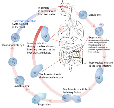
Entamoeba histolytica Updated: 01/26/2024 © Jun Wang, MD, PhD   General features A protozoan transmitted by ingestion of amebic cysts   Fecal-oral transmission usually through food or water Most cases asymptomatic, while up to 100k death annually  Life cycle Key clinical features Intestinal amebiasis and extraintestinal manifestations Bloody diarrhea due to colonic tissue damage Presentation depends on locations of infection Gastrointestinal: Gradual onset, fever, abdominal pain, tenesmus, diarrhea (with or without blood), dysentery Liver: Abscess, most common extraintestinal complication; fever, right upper quadrant pain, hepatomegaly with hepatic tenderness; may rupture and involve surrounding structures Respiratory tract: Rare, atelectasis and pleural effusions Brain: Abscesses, very rare, sudden onset symptoms such as headache, vomiting, and mental status changes with rapid progression to death Colonoscopic

Bacterial gastroenteritis

Bacterial gastroenteritis Updated: 01/26/2024 © Jun Wang, MD, PhD   General features Infection of bacteria Produce endotoxin and exotoxin Secretory Bacillus cereus: rice Enterotoxigenic E. coli : recent travel Clostridium perfringens: undercooked meat, raw legumes Vibrio cholerae : Sea food, water Staphylococcus aureus: Inadequately refrigerated food Invasive Yersinia: mile, pork Typhi or paratyphi salmonella: recent travel Inflammatory Campylobacter : recent travel Enterohemorrhagic E. coli : meat, may cause HUS Clostridium difficile : antibiotics Shigella : recent travel, may cause HUS Noncholera vibrio: shellfish Nontypoidal salmonella : poultry and eggs Endotoxin Components of the cell wall of G-negative bacteria Released by death of bacterial, antibiotics, or antibodies Cause fever: Release of interleukin-1 Cause shock : TNF associated hypotension and endothelial damage Release of nitric oxide: Vasodilation, bacterial crossing blood-brain barrier Ex

Escherichia coli

Image
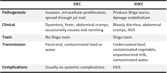
Escherichia coli Updated: 01/30/2024 © Jun Wang, MD, PhD   General features Anaerobic G- bacteria, part of normal intestinal flora Most common cause of bacterial diarrhea worldwide Infections due to disruption of the mucosa Most ferment sorbitol (dark pink colonies), except EHEC O157:H7 (white colonies), on MacConkey agar 5 commonly seen diarrhea-associated groups ETEC : Enterotoxigenic E coli, watery diarrhea EIEC : Enteroinvasive E coli, dysentery EHEC : Enterohemorrhagic E coli, hemorrhagic colitis and hemolytic uremic syndrome EPEC : Enteropathogenic E coli, infantile diarrhea EAEC : Enteroaggregative E coli, persistent diarrhea in children and patients infected with HIV Enterotoxigenic E. coli Major cause of “traveler’s diarrhea” and diarrhea in <3-year-olds in developing countries Bind to small intestine epithelium through colonization factors (CFs) Capsule resistant to phagocytosis Two enterotoxins, detectable by immunoassay, etc LT: heat-labile, activate

Norovirus/Norwalk virus

Norovirus/Norwalk virus Updated: 01/29/2024 © Jun Wang, MD, PhD   General features Most common cause of epidemic nonbacterial gastroenteritis Norovirus   Single-stranded RNA and hexagonal capsid 5 genogroups, I, II and IV are human pathogens Highly contagious, Fecal-oral transmission Usually not severely ill Lasting 24-72 hours, usually self-limited Associated with shellfish, prepared foods, etc Diarrhea due to transient malabsorption of D-xylose and fat and inhibition of brush-border enzymes including alkaline phosphatase and trehalase Key clinical features Usually not severely ill Nausea, vomiting, moderate watery diarrhea (4-8 stools/day) Signs of volume depletion: tachycardia, hypotension, ONLY in severe cases Stools usually do NOT contain mucous or leukocytes Diagnosis Usually clinical diagnose Confirmation needed for immunocompromised patients with severe or persistent symptoms Multi-pathogen molecular tests to confirm Other pathogens need to be ruled out

Infectious gastroenteritis

Image
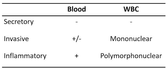
Infectious gastroenteritis Updated: 01/26/2024 © Jun Wang, MD, PhD   General features Inflammation of GI tract Most commonly caused by viruses May be associated with bacteria, fungi or parasites Fecal-oral transmission: foodborne, waterborne Most important risk factor: poor hygiene and sanitation Presentations: abdominal pain, nausea/vomiting, diarrhea Mild cases Most common Likely self-limited Supportive therapy only Severe cases May cause dehydration and sepsis   Diarrhea vs. Dysentery Diarrhea o    Passage of loose or watery stools o    At least 3 times in a 24-hour period o    Increased water content due to impaired water absorption or active water secretion, with or without mucosa injury or inflammation o    Acute: < 14 days; Persistent: 14-30 days; Chronic: > 30 days Dysentery o    AKA invasive diarrhea o    Diarrhea with visible blood or mucus o    Commonly associated with fever and abdominal pain   Types of diarrhea Secretory o