Posts

Showing posts with the label Oral cavity

Practice question answers oral cavity pathology

Practice question answers, oral cavity pathology Updated: 03/07/2019 © Jun Wang, MD, PhD 1. B. Polypoid growth with fibrous core and smooth non-neoplastic squamous covering is most compatible with irritation fibroma , a reactive process. Erythroplakia is usually a reddish patch/plaque that commonly harbors dysplasia. Pyogenic granuloma is indeed a capillary hemangioma with lobulated growth. Squamous cell carcinoma has irregular growth, invasion and cytological atypia. Squamous papilloma has finger like projects with fibrovascular core and benign squamous covering. 2. A. A small shallow oral erosion/ulcer without other presentations is most likely aphthous ulcer . Erythroplakia is usually a reddish patch/plaque that commonly harbors dysplasia. Hairy leukoplakia is a white patch/plaque with a smooth or “hairy” surface. Herpes has clusters of small painful vesicles. Squamous cell carcinoma has mass, with or without ulceration. 3. D. Herpes has clusters of small pai
Practice questions Oral cavity pathology

Image
Practice questions Oral cavity pathology Updated: 02/28/2019 © Jun Wang, MD, PhD 1. A 35-year-old man presents with a firm left gum nodule for 3 months. His past medical history is unremarkable. He has a 15 pack-year history of cigarette smoking. He does not drink alcohol. Physical examination reveals a 0.7 cm firm polypoid mass with focal erosion and exudates. The mass is removed. Microscopically it has a fibrous core with slightly hyperplastic smooth squamous lining. The squamous epithelium has parakeratosis and mild cytological atypia. What is the diagnosis? A. Erythroplakia B. Irritation fibroma C. Pyogenic granuloma D. Squamous cell carcinoma E. Squamous papilloma 2. A 15-year-old boy presents with painful lesion of his left lateral tongue. He has a history of infectious mononucleosis a year ago. Physical examination reveals an isolated 0.2 cm shallow ulcer at the left anterolateral tongue. The ulcer has well-defined erythematous edge. No other abnormalities

Oral squamous cell carcinoma

Oral squamous cell carcinoma   Updated: 02/12/2021 © Jun Wang, MD, PhD General features Majority of oral cavity cancers Usually ages 50-70 years Predominantly men Likely second primary tumors due to chronic exposure to carcinogens prior ( field cancerization ) Most tonsil area cases are HPV associated HPV associated cancer tends to have better prognosis Risk factors Major: Alcohol, tobacco, chewing tobacco, marijuana, betel nuts and pan (India) Other: HPV, EBV, etc Sunlight exposure for lip cancer Fruit and vegetable consumption significantly reduces risk Clinical presentations Leukoplakia: smooth or warty white patch/plaque, cannot be scraped off Erythroplakia : Red area, more likely have squamous dysplasia Mass Ulcer Key morphological features Atypical cells with squamous differentiation ( intercellular bridges and squamous pearls ) Treatment Surgery Radiation therapy May consider photodynamic and chemotherapy Back to oral cavity

Odontogenic diseases

Odontogenic diseases   Updated: 02/12/2021 © Jun Wang, MD, PhD Odontogenic cysts Derived from remnants of odontogenic epithelium Within the maxilla or mandible or surrounding soft tissue Heterogenous group of lesions Inflammatory: Periapical/radicular cyst, most common, tooth bearing areas; squamous epithelium lined tissue with variable inflammation Developmental: Lateral periodontal cyst Neoplastic: Keratocystic odontogenic tumor Odontogenic tumors Rare Derived from remnants of odontogenic epithelium Present within the maxilla or mandible or surrounding soft tissue Either neoplastic (benign or malignant) or hamartomatous Back to oral cavity pathology Back to contents

Leukoplakia and erythroplakia

Leukoplakia/erythroplakia   Updated: 02/12/2021 © Jun Wang, MD, PhD General features Hyperkeratotic patches of clonal squamous cell growth More common in old male Associated with tobacco use, alcohol, ill-fitting dentures, chronic irritants  HPV less likely important May be precancerous, higher risk if in floor of mouth or ventral surface of tongue Clinical term, considered precancerous until proven otherwise May have spontaneous regression Clinical presentations Leukoplakia: smooth or warty white patch/plaque, cannot be scraped off Erythroplakia : Red area, more likely have squamous dysplasia Key morphological features Hyperplastic oral mucosa with squamous dysplasia (loss of maturation) Treatment Immediate biopsy if change in appearance Surgery, laser ablation, cryosurgery, retinoids, etc Back to oral cavity pathology Back to contents

Nonspecific inflammatory disorders of oral cavity

Image
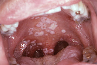
Nonspecific inflammatory disorders of oral cavity   Updated: 02/12/2021 © Jun Wang, MD, PhD Gingivitis Usually due to poor oral hygiene May be associated with scurvy, endocrine alterations (teenagers, pregnancy), disseminated tuberculosis, phenytoin or other drugs Painless inflammation of gingiva around gingival sulci or cuff surrounding each tooth, due to bacterial plaque Dental plaque : Mixture of proteinaceous debris and bacteria, may be calcified Periodontitis Alveolar bone destruction May lead to tooth loss if progressive Due to recurrent and continuous gingivitis  Pockets around teeth > 3 mm in depth Management: Surgery, brushing and flossing Aphthous ulcer Common, especially in people younger than 20 Painful, recurrent  Shallow small ulcer with well defined border Unknown etiology and pathogenesis May be associated with other immune disorders Clinical diagnosis Usually resolve spontaneously Irritation fibroma Reactive process Due to chr

Hairy leukoplakia

Hairy leukoplakia   Updated: 02/12/2021 © Jun Wang, MD, PhD General features Lateral border of tongue in immunocompromised patients Associated with HIV+ patients but caused by Epstein-Barr virus No specific treatment needed Clinical presentations Adherent white patches with smooth or irregular "hairy" surface Key morphological features Hyperkeratosis and parakeratosis Balloon cells Treatment Focus on underlying conditions Back to oral cavity pathology Back to contents

Pathology of oral cavity

Pathology of oral cavity   Updated: 02/20/19 © Jun Wang, MD, PhD Key anatomy and histological features Squamous epithelium Minor salivary glands Nonspecific inflammatory disorders Pyogenic granuloma Squamous abnormalities Hairy leukoplakia Leukoplakia/erythroplakia Squamous cell carcinoma Odontogenic disorders Odontogenic cyst Odontogenic tumors Practice questions Back to contents