Posts

Showing posts with the label breast

Usual ductal hyperplasia

Usual ductal hyperplasia Updated: 12/10/2018 © Jun Wang, MD, PhD General features AKA papillomatosis Slightly increased risk of subsequent malignancy Mild to florid hyperplasia, based on degree of ductal proliferation Slightly increased risk for invasive carcinoma if moderate to florid hyperplasia Clinical presentations Mammogram abnormalities Mass Key morphological features Increased number of epithelial cells within preexisting glandular components Mild hyperplasia: 2-4 epithelial layers Moderate hyperplasia: 4 or more epithelial layers Florid hyperplasia : Epithelium almost completely fills duct but with fenestrations (irregular lumen at periphery) and papillomatosis No increased number of glands (not adenosis) NO cytological atypia Marker Positive for keratin 903 (negative in atypical ductal hyperplasia) Treatment Usually no need for treatment Follow up Back to breast pathology Back to contents
Sclerosing adenosis

Sclerosing adenosis Updated: 12/10/2018 © Jun Wang, MD, PhD General features Glandular and stromal proliferation Mean age 30 years Slightly increased risk for invasive carcinoma Clinical presentations Recurring pain that tends to be linked to the menstrual cycle Mammogram abnormalities Key morphological features Increased number of distorted/compressed small glands Background stromal proliferation/fibrosis NO cytological atypia Intact myoepithelial cell layer (marked by positive reactivity to CD10, actin, p63) Treatment Usually no treatment needed Follow up Back to breast pathology Back to contents

Radial scar

Radial scar Updated:12/10/2018 © Jun Wang, MD, PhD General features Benign Mimic invasive carcinoma grossly and histologically Slightly increased risk for invasive carcinoma , due to coexisting proliferative disease Clinical presentations Mammogram abnormalities Key morphological features Stellate arrangement of compressed ducts Central sclerosis and elastosis NO cytological atypia Intact myoepithelial cell layer (marked by positive reactivity to CD10, p63) Treatment Observation if small Excision if large, or malignancy suspected Back to breast pathology Back to contents

Phyllodes tumor

Phyllodes tumor Updated:07/01/2023 © Jun Wang, MD, PhD General features Biphasic tumor with hypercellular stroma Three categories based on histology: benign, borderline and malignant Malignant type more common in older women Local recurrence common if not completely removed Malignant type may metastasize to lung, bone, CNS 1q+ most common chromosomal abnormality Clinical presentations Firm mass Key morphological features Leaf like processes Hypercellular stroma Variable hemorrhage, necrosis Treatment Excision Back to breast pathology Back to contents

Invasive carcinoma breast

Invasive carcinoma of breast Updated: 12/11/2018 © Jun Wang, MD, PhD General features More common in upper outer quadrant Classified according to architecture and cytological grade Most common type: Ductal carcinoma, followed by lobular carcinoma Arises from terminal duct lobular unit Always need ancillary test (ER, PR, Her2) Clinical presentations Mass Nipple abnormalities Mammographic detection of microcalcifications, soft-tissue densities, or both Key morphological features Mass with infiltrating border Hemorrhage and/or necrosis NO myoepithelial cells (can be stained by CD10 and p63) Invasive ductal carcinoma With better tubule formation : irregular tubules lined by atypical cells With poor tubule formation : less tubular structure Positive for E-cadherin Invasive lobular carcinoma Commonly bilateral and multifocal Relatively monotonous tumor cells Single files , no tubular formation May have intracellular mucin, signet ringcells N

Intraductal papilloma

Intraductal papilloma Updated: 12/10/2018 © Jun Wang, MD, PhD General features Benign Intraductal proliferation of epithelial and myoepithelial cells Usually mid age Clinical presentations Bloody nipple discharge Key morphological features Fibrovascular connective tissue core Covered by epithelial and myoepithelial cells NO cytological atypia Intact myoepithelial cell layer (marked by positive reactivity to CD10, p63) Treatment Observation if small Excision if large, or presence of atypia Excision if found on core biopsy Back to breast pathology Back to contents

Gynecomastia

Gynecomastia Updated: 07/01/2023 © Jun Wang,MD, PhD General features Most common lesion of male breast Usually bilateral, but may be more distinct in one breast Caused by increase in estrogen to androgen ratio Multifactorial etiology: Physiological (puberty or aging), endocrine tumors, endocrine dysfunctions, non-endocrine diseases, drug induced, idiopathic May regress without treatment Clinical presentations Button or disk-like subareolar enlargement Key morphological features Hypertrophy and hyperplasia of glands and stroma NO cytological atypia Intact myoepithelial cell layer (marked by positive reactivity to CD10, p63) Treatment Observation Treatment of underlying causes Excision if not regress Back to breast pathology Back to contents

Fibrocystic changes

Fibrocystic changes Updated: 12/10/2018 © Jun Wang,MD, PhD General features A general category for various entities Cystic features, with or without fibrosis Commonly ages 25-45 years Either proliferative (adenosis, hyperplasia) or nonproliferative (cysts) If prolifereative, slightly increased risk of cancer development Pathogenesis Associated with estrogen effects Clinical presentations Usually multifocal and bilateral Breast pain that worsens during ovulation Tender nodular swellings Key morphological features Cysts formation with or without following Fibrosis Increased number of glands Apocrine metaplasia (more eosinophilic cytoplasm) Calcification NO cytological atypia Treatment Aspiration Surgery Back to breast pathology Back to contents

Fibroadenoma

Fibroadenoma Updated: 12/11/2018 © Jun Wang,MD, PhD General features Biphasic tumor with epithelial and stromal components Most common benign tumor of female breast Usually young women Hormonally responsive, grows during pregnancy and late luteal phase, regresses after menopause Slightly increased risk of carcinoma, especially when ductal hyperplasia presents Clinical presentations Well demarcated mobile firm mass Key morphological features Sharply circumscribed with smooth, rounded border Stromal proliferation with stretched and compressed glands/ducts Treatment Excision Back to breast pathology Back to contents

Ductal ectasia

Ductal ectasia Updated: 12/10/2018 © Jun Wang, MD, PhD General features Usually women in reproductive years Occasionally in neonates Associated with smoking, possibly pituitary adenomas and increased prolactin levels Clinical presentations Painful, erythematous, subareolar mass Nipple discharge Key morphological features Dilated large ducts Fibrous thickening of wall Foamy macrophages and proteinaceous debris in lumen May have calcification Treatment Aspiration/Drainage Surgery Back to breast pathology Back to contents

Breast Paget disease

Breast Paget disease Updated:02/24/2020 © Jun Wang, MD, PhD General features Arises in or involves the main excretory ducts Associated with underlying malignancy, unlike extramammary Paget disease Clinical presentations Eczema like lesion involving nipple/areola Key morphological features Pale malignant epithelial cells randomly disposed within epidermis of nipple/areola Differential diagnosis Melanoma: Positive for S-100, HMB45, negative for cytokeratin Paget disease: Negative for S-100 and HMB45, positive for cytokeratin (CAM5.2, CK7) Treatment Nipple removal as part of treatment of underlying malignancy Back to breast carcinoma Back to breast pathology Back to contents

Inflammatory breast diseases

Inflammatory diseases of breast Updated: 04/10/2023 © Jun Wang, MD, PhD Acute mastitis/abscess Infection or non-infection associated inflammation Usually bacterial if infected Associated with lactation and cracks in nipple Pregnancy related infections: Staphylococcus, unilateral, often MRSA Zuska’s disease Periareolar abscess associated with squamous metaplasia of lactiferous ducts May form fistula Clinical features: Acute inflammation: fever, pain, erythematous changes, swelling Pathological features: diffuse neutrophilic infiltrate Treatment Antibiotics Incision and drainage, if abscess formed Lymphocytic mastopathy AKA diabetic mastopathy More commonly associated with type I diabetes May occur in type II diabetes or non-diabetes, or associated with autoimmune disorders Clinical features: Palpable mass , usually subareolar Pathological features: Dense polyclonal lymphocytic infiltrates, stromal fibrosis Treatment: Usually NOT needed Surger

Carcinoma in situ

Breast carcinoma in situ Updated: 12/11/2018 © Jun Wang, MD, PhD General features Neoplastic proliferation with malignant features Confined within spaces bordered by myoepithelium and basement membrane May progress to invasive carcinoma Clinical presentations Clinically occult Ductal carcinoma in situ Mammographic finding of microcalcifications, soft-tissue densities Lobular carcinoma in situ:  Usually incidental finding due to biopsy for other lesions, such as fibrocystic changes Multifocal, bilateral NOT associated with microcalcification or stromal density Key morphological features Classified according to architecture and cytological grade Ductal carcinoma in situ Comedo type : Markedly atypical ductal cells with central necrosis, may habor microinvasion, may be treated as invasive ductal carcinoma Paget disease, ductal carcinoma in situ involving nipple skin Cribriform : Round rigid spaces (Roman bridges) Micropapillary : finger like proj

Atypical ductal hyperplasia

Atypical ductal hyperplasia Updated: 12/10/2018 © Jun Wang, MD, PhD General features Neoplastic intraductal lesion with architectural and cytological features suggestive but not diagnostic of ductal carcinoma in situ May be multicentric Higher risk of carcinoma Clinical presentations No specific clinical features Microcalcification/architecture changes in mammogram Key morphological features Less than 2-3 mm, if larger, diagnosed as ductal carcinoma in situ Micropapillae, tufts, bridges, solid and cribriform patterns Monomorphic cells with rounded or ovoid nuclei NO cytological atypia Intact myoepithelial cell layer (marked by positive reactivity to CD10, p63) Marker Negative for keratin 903 (positive in usual ductal hyperplasia) Treatment Surgery to rule out accompanying carcinoma Follow up Back to breast pathology Back to contents

Breast carcinoma

Image
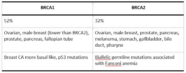
Breast carcinoma Updated: 02/14/2020 © Jun Wang, MD, PhD General feature Most common cancer in women, regardless of race or ethnicity Second most common cause of death from cancer in women Mortality reducing due to early detection by mammogram Etiology Genetic Hormonal: most commonly associated risk factor Higher risk group Age: risk increases with age until age 80 Personal history of breast cancer Inherited genetic mutations for breast cancer History of atypical hyperplasia Mammographically dense breasts Intermediate risk group High endogenous estrogen or testosterone levels, etc Carcinogenesis Estrogen dependent: most common, ER+ Her2 associated: most common in Li-Fraumeni syndrome ER, Her2 independent Molecular abnormalities Hereditary Breast and Ovary Cancer syndrome: BRCA1 and BRCA2 Her2 p53 CHEK2 PTEN: Cowden syndrome STK11, as seen in Peutz-Jeghers syndrome ATM CDH1 in lobular carcinoma DNA mismatch repair genes (MLH1

Pathology of breast

Pathology of breast Updated:01/12/2019 © Jun Wang, MD, PhD Anatomy/histology Specialized sweat glands Suspensory ligaments : linking skin to fascia of pectoralis major and pectoralis minor Lymphatic drainage Axillary lymph nodes: majority of drainage Internal thoracic lymph nodes Posterior intercostal Supraclavicular nodes Terminal duct lobular unit Glandular cells: secretion Myoepithelial cells: assist in mild ejection, absent in invasive carcinoma Clinical presentations Pain: physiological or pathological, usually due to inflammation if pathological Mass: benign or malignant Nipple discharge Galactorrhea: Drugs, hyperprolactinemia (pituitary tumor), etc Bloody discharge: Probably intraductal papilloma Other discharge: Ductal ectasia, fibrocystic changes, neoplasms Non-neoplastic breast lesions Inflammatory breast diseases Acute mastitis/abscess Lymphocytic mastopathy Fat necrosis Idiopathic granulomatous mastitis Ductal ectasia