Posts

Showing posts with the label pancreas

Serous cystic neoplasm

Serous cystic neoplasm Updated: 03/26/2019 © Jun Wang, MD, PhD General features Also called serous cystadenoma, microcystic adenoma, glycogen rich cystadenoma Benign, more common in women, mean age 66 May be associated with von Hippel Lindau syndrome Clinical presentations More common in pancreatic body or tail Local discomfort/pain, or pancreatic obstruction if in pancreatic head May be asymptomatic Key pathological features Multiloculated mass , sharply outlined, filed with clear fluid Small cystic spaces lined by small cuboidal cells with clear cytoplasm (glycogen) Markers Positive for cytokeratin, inhibin Treatment Surgery Back to exocrine pancreas pathology Back to contents
Solid-pseudopapillary neoplasm

Solid-pseudopapillary neoplasm Updated: 03/26/2019 © Jun Wang, MD, PhD General features AKA papillary and solid epithelial neoplasm, papillary cystic neoplasm, Gruber-Frantz tumor Low grade malignancy Commonly 30-35 year old women, not associated with any clinical syndrome Poor prognostic factors: Vascular invasion, high nuclear grade, “necrobiotic nests” Metastases in 10-15% Clinical presentations Epigastric pain, nausea, vomiting, weight loss, etc Key pathological features Poorly cohesive monomorphic epithelial cells forming solid and pseudopapillary structures Pseudopapillae with fibrovascular cores Several layers of bland fragile epithelial cells with clear to eosinophilic cytoplasm Cysts formed due to solid nests minus cells degenerating away from the small vessels Molecular abnormality Gain-of-function mutation of beta-catenin Treatment Surgery Back to exocrine pancreas pathology Back to contents

Non neoplastic pancreatic cysts

Non neoplastic pancreatic cysts Updated: 03/26/2019 © Jun Wang, MD, PhD Pseudocyst Painful, collections of pancreatic secretions after pancreatitis ( acute or chronic ), trauma, ductal calculi, obstructive neoplasms May be asymptomatic Cysts without epithelial lining Complications: abscess, erosion into blood vessels, hemorrhage, infection, perforation into a hollow viscus Management: follow up with image studies, supportive care, drainage if symptomatic Congenital cysts Commonly multiple No communication with duct system Often associated with polycystic disease affecting kidney and liver and von Hippel Lindau syndrome Lined by a single layer of epithelium with a fibrous wall Back to exocrine pancreas pathology Back to contents

Mucinous cystic neoplasm

Mucinous cystic neoplasm   Updated: 03/26/2019 © Jun Wang, MD, PhD General features Benign or potentially low grade malignant Cystic epithelial neoplasm composed of mucin producing cells One of three precursor lesions of pancreatic adenocarcinoma Almost always women, mean age 45 years May be associated with invasive carcinoma Metastases usually restricted to abdominal cavity Need to differentiated from primary ovarian mucinous neoplasm if ovarian involved Clinical presentations Abdominal pain, recurrent pancreatitis, gastric outlet obstruction, and/or mass Jaundice and/or weight loss if malignant Key pathological features Usually in body/tail, multilocular (occasionally unilocular) megacysts No communication with ductal system unless fistula formed Cyst wall papillary, trabecular or thickened Mucoid/watery cyst contents Mucinous epithelium with dense “ ovarian type ” stroma Ovarian type stroma is ER+, PR+, inhibin+, CD10+ May present with low- or int

Intraductal papillary mucinous neoplasm

Intraductal papillary mucinous neoplasm Updated: 03/26/2019 © Jun Wang, MD, PhD General features Intraductal epithelial neoplasm of mucin producing cells Arising in main pancreatic duct or its branches Benign or malignant Classified based on highest degree of cytoarchitectural atypia and invasiveness More common in men age 60+ at head of pancreas Risk factor for pancreatic cancer Associated with Peutz-Jegher syndrome Clinical presentations Epigastric pain, weight loss, jaundice, diabetes, pancreatitis Key pathological features Papillary growth of mucinous cells with duct dilatation (cyst formation), and dysplasia Main duct involvement : Usually diffusely dilated, tortuous and irregular duct filled with mucin; higher risk of high grade dysplasia and malignancy Branch duct involvement : Multicystic, grape-like structures filled with tenacious mucin; cyst walls usually thin with flat or papillary lining; cysts separated by normal pancreas Treatment Sur

Chronic cholecystitis

Chronic cholecystitis Updated: 03/26/2019 © Jun Wang, MD, PhD General features Very common Most in women, 40+ Majority associated with cholelithiasis Complications: Acute cholecystitis , acute pancreatitis , biliary fistulas, choledocholithiasis, gallstone ileus Usually associated with bacteria and other microorganisms Clinical presentations May be asymptomatic Recurrent acute cholecystitis or biliary colic Pathological features Variable thickening of gallbladder wall, variable adhesions with chronic inflammation Porcelaingallbladder : Dense, paucicellular hyaline fibrosis with diffusely effacements of normal structure Calcifications Higher risk for gallbladder carcinoma Management Cholecystectomy Back to exocrine pancreas pathology Back to contents

Chronic pancreatitis

Image
Chronic pancreatitis Updated: 03/25/2019 © Jun Wang, MD, PhD General features Continuing, chronic, inflammatory process Irreversible loss of pancreatic parenchyma and replacement with fibrosis Variable pancreatic insufficiency (malabsorption, diabetes) More common in men, 40+ years of age, alcoholics May simulate or coexist with pancreatic carcinoma Complications: Pseudocysts; widespread fat necrosis (from liberation of lipase), localized portal hypertension due to fibrosis of splenic vein in alcoholic hepatitis Etiology Commonly idiopathic Alcohol : most common etiology (60-70%) Intraductal plugging and obstruction : Stones, tumors Toxins and toxic metabolites : Promotes release of cytokines Oxidative stress: Idiopathic pancreatitis Necrosis-fibrosis: Recurrent acute pancreatitis that heals with fibrosis Ischemia: From obstruction and fibrosis; important in exacerbating disease Pathogenesis Destruction of pancreatic tissue due to recurrent inflammation

Pancreatic cancer

Image
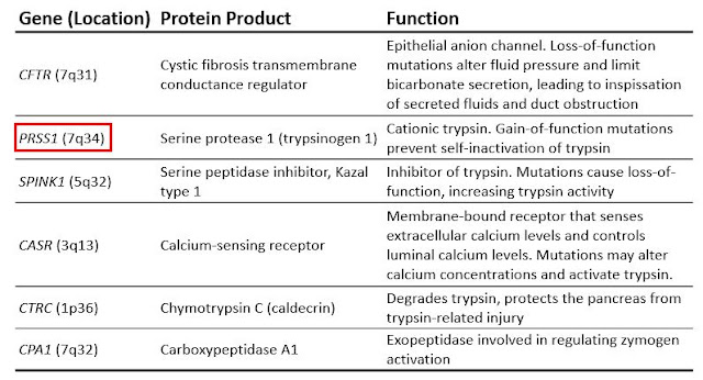
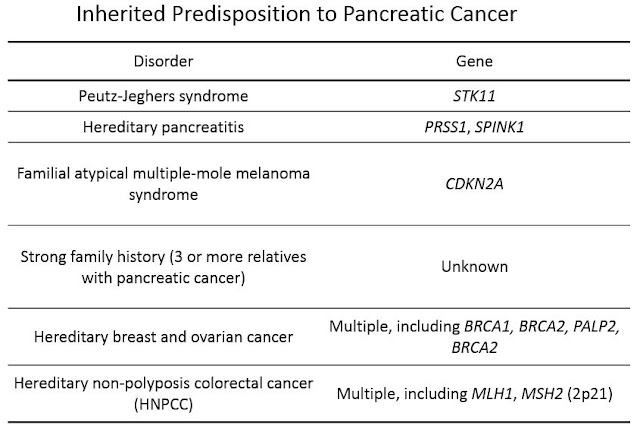
Pancreatic cancer Updated: 03/02/2021 © Jun Wang, MD, PhD General features Classification based on differentiation : ductal, endocrine , acinar, etc Majority are ductal carcinoma Fourth leading cause of death More common in population older than 50 3 recognized precursors of invasive disease: PanIN , IPMN and mucinous cystic neoplasm Most common in head and body Overall poor prognosis Clinical presentations Non specific: Anorexia, malaise, nausea, fatigue, and midepigastric or back pain Painless obstructive jaundice: Most characteristic sign of cancer of head of the pancreas Risk factors Smoking, obesity, diabetes mellitus , chronic pancreatitis Genetic abnormalities Molecular abnormality KRAS, CDKN2A, p53, SMAD4, BRCA2, SMAD4, etc Inherited Ductal adenocarcinoma Most common pancreatic cancer Non specific presentation including pain, weight loss, anorexia, malaise, weakness Trousseau sign : Migratory thrombophlebitis, due to tumor or tumor

Cholelithiasis

Cholelithiasis   Updated: 03/16/2021 © Jun Wang, MD, PhD General features Affects 10% of adults in developed countries Cholesterol stones: Most common Risk factors: “ Fat, fertile [multiple pregnancies], forty, female ”; rapid weight loss, certain genetic disorders, low HDL levels Pigment stone risk factors: Increased unconjugated bilirubin Clinical presentations May be asymptomatic Acute or chronic cholecystitis , choledocholithiasis, cholangitis, gallstone ileus, acute pancreatitis Diagnosis Ultrasound X-ray if contains calcium Management Cholecystectomy Back to exocrine gallbladder pathology Back to contents

Autoimmune pancreatitis

Autoimmune pancreatitis   Updated: 02/13/2021 © Jun Wang, MD, PhD General features AKA primary sclerosing cholangitis-like pancreatitis, non-alcoholic duct destructive chronic pancreatitis Two subtypes Lymphoplasmacytic sclerosing autoimmune pancreatitis Idiopathic duct centric autoimmune pancreatitis Forms mass that may constrict bile duct Clinically and radiological features may mimic malignant tumors May cause exocrine and endocrine deficiencies Risk factor for pancreatic cancer Overall excellent prognosis Lymphoplasmacytic sclerosing autoimmune pancreatitis IgG4 related systemic disease More common in male , older patients , often presents with jaundice Associated with sclerosing cholangitis, IgG4 related dacryoadenitis and sialadenitis , Sjögren-like syndro me, retroperitoneal fibrosis , obliterative phlebitis, arterial involvement Idiopathic duct centric autoimmune pancreatitis No gender preference, younger patients, associated with inflammato

Acute pancreatitis

Image
Acute pancreatitis Updated: 03/25/2019 © Jun Wang, MD, PhD General features Clinical emergency More common in women, in African Americans Commonly associated with biliary tract disease in women , alcoholism in men Overall mortality: 10% if edematous vs. 50% if hemorrhagic/necrotic, higher in biliary pancreatitis , most deaths due to failure of multiple organ system in the first week, and infections in subsequent weeks Etiology Metabolic: Alcoholism ; hyperlipoproteinemia, hypercalcemia; certain drugs (e.g., azathioprine); cigarette smoking Genetic: Mutation of trypsin , trypsin regulators , or calcium metabolism regulators Mechanical: Bile duct obstruction (increase intrapancreatic ductal pressure and leads to the accumulation of enzyme-rich fluid in the interstitium); trauma; iatrogenic injury including perative injury/Endoscopic procedures with dye injection Toxins: Such as scorpion bite in Trinidad Vascular: Shock; atheroembolism; vasculitis Infectious: Mu

Acute cholecystitis

Acute cholecystitis   Updated: 03/16/2021 © Jun Wang, MD, PhD General features With gallstone (acute calculous cholecystitis, >90% cases) or not (acute acalculous cholecystitis) Associated with obstruction, biliary colic, ischemia and bacterial infection May results in perforation Clinical presentations Acute inflammation: abdominal pain, right upper quadrant tenderness, nausea, vomiting, fever, leukocytosis, mild jaundice Murphy sign Pathological features Enlarged, distended gallbladder Acute inflammation, serosal and mucosal exudate, hemorrhage and necrosis Laboratory features Leukocytosis Serum bilirubin and alkaline phosphatase may be NORMAL unless bile duct obstructed Sonographic findings Gallbladder wall thickening or edema (double wall sign) Stones Acute gangrenous cholecystitis Mural infarction High risk of perforation Associated with Clostridium perfringes and air in gallbladder (pneumobilia) High mortality rate Transmural inflammat

Pathology of exocrine pancreas and gallbladder

Pathology of exocrine pancreas and gallbladder Updated: 03/14/19 © Jun Wang, MD, PhD Key anatomy and histological features Head, body and tail  Exocrine pancreas: Majority of pancreatic tissue, glandular epithelial cells Predominantly at the head Drain through pancreatic duct Acini, ducts Endocrine pancreas: Islets, pale endocrine cells Alpha: secrete glucagon (20% 0f cells) Beta: insulin (70% of cells) Delta: somatostatin (5% of cells) PP cells: pancreatic polypeptide Non neoplastic disorders Acute pancreatitis Chronic pancreatitis Autoimmune pancreatitis Cysts Cystic neoplasm Serous cystic neoplasm Mucinous cystic neoplasm Intraductal papillary mucinous neoplasm Solid-pseudopapillary neoplasm Non cystic neoplasm Pancreatic cancers Gallbladder disorders Acute cholecystitis Chronic cholecystitis Cholelithiasis Malignant tumor of gallbladder Practice questions Back to gastrointestinal tract pathology Back to conte

Practice questions answers II Pathology of endocrine pancreas

Practice questions answers Pathology of endocrine pancreas II © Jun Wang, MD, PhD 1. E. Dizziness and sweating are commonly seen in patient with hypoglycemia , the most common acute complication in patient with diabetes . It can be confirmed with abnormally low blood glucose. Diabetic ketoacidosis has lower arterial pH and elevated ketone in urine, as well as hyperglycemia. Diabetic nephropathy has proteinuria and is more likely a chronic process without sudden onset of neurological symptoms, and does not cause hypoglycemia . Diabetic neuropathy is a chronic process involving both motor and sensory functions, but does not cause hypoglycemia . Hyperosmolar hyperosmotic syndrome has a blood glucose > 600 mg/dl. 2. D. The most common causes of hypoglycemia in patients with diabetes include skipping meals, excessive exercise, excess insulin administration, etc. Excess fatty acid metabolism is seen in diabetic ketoacidosis . Glucagonoma causes hyperglycemia, not hypo

Practice questions II Pathology of endocrine pancreas

Practice questions Pathology of endocrine pancreas II © Jun Wang, MD, PhD 1. Use this case for the next two questions . A 59-year-old man presents with dizziness and sweating for 2 hours. He was diagnosed with type 2 diabetes two months ago, and has been compliant with his medication. In addition, he started an aggressive diet plan to reduce weight. He skipped his breakfast and hiked 3 miles before he felt dizzy. His past medical history include hypertension and obesity. He does not drink alcohol or smoke cigarette. Physical examination reveals a BMI at 35 (normal 18-25). He appears to be slightly confused and his skin is moist. His heart rate is 135 bpm. No other abnormalities are noted. Laboratory test reveal a glucose at 52 mg/dl (normal 70-100 mg/dl) and HbA1C at 8.0% (normal 4-5.6%). Other laboratory test results are within normal range. What is the diagnosis? A. Diabetic ketoacidosis B. Diabetic nephropathy C. Diabetic neuropathy D. Hyperosmolar hyperosmotic syn