Posts

Pulmonary hypertension

Pulmonary hypertension Updated: 10/07/2022 © Jun Wang, MD, PhD   Definition Elevated pulmonary arterial pressure ≥ 25 mm Hg (or 20 mm Hg + Pulmonary vascular resistance ≥ 3 Woods units, debating), or   > 30 mm Hg during exercise Severe pulmonary hypertension mPAP is ≥ 35 mm Hg, or mPAP is > 20 mm Hg with an elevated right atrial pressure (>14 mm Hg) and/or the cardiac index is <2 L/min/m 2 Precapillary pulmonary hypertension: Due to pulmonary Caused by pulmonary artery remodeling Low pulmonary artery wedge pressure Post-capillary pulmonary hypertension Caused by elevated pulmonary vein pressure, due to left heart dysfunction High pulmonary artery wedge pressure WHO classification Group 1: Pulmonary arterial hypertension (PAH) Group 2: Pulmonary hypertension due to left-sided heart disease Group 3: Pulmonary hypertension due to lung diseases and/or hypoxia Group 4: Pulmonary hypertension due to pulmonary artery obstruction Group 5: Pulmonary hypertension
Lymphangioleiomyomatosis

Lymphangioleiomyomatosis Updated: 11/01/2021 © Jun Wang, MD, PhD   General features Cystic destruction with progressive dysfunction of lung Proliferation of lymphatic smooth muscle-like cells Probably originated from perivascular epithelioid cell Most common in reproductive age women Associated with tuberous sclerosis Key clinical features Most common presentations: Dyspnea, cough, pleural effusion, etc Pneumothorax commonly seen Key morphological features Cystic changes Proliferation of spindle cells (LAM cells), may be nodular Genetic abnormalities TSC1 and TSC2 Diagnosis Radiologic studies: Cystic changes Pathological identification of LAM cells Markers Positive: HMB45 Treatment Treatments of complications Lung function improvement Lung transplantation   Back to respiratory tract neoplasms Back to contents  

Renal medullary carcinoma

Renal medullary carcinoma Updated: 05/05/2021 © Jun Wang, MD, PhD General features Aggressive Predominantly seen in patients with sickle cell disorder More common in male, mean age 26 Loss of SMARCB1/INI1 Pathological features Markedly pleomorphic tumor cells Various pattern Back to renal cell carcinoma Back to kidney masses Back to contents

MUTYH-associated polyposis

MUTYH-associated polyposis   Updated: 03/02/2021 © Jun Wang, MD, PhD General features AKA MYH-associated polyposis Autosomal recessive Considered a mild form of familial adenomatous polyposis Biallelic mutation of MUTYH gene (DNA mismatch repair) Less 1% of patients with colorectal cancer Pathogenesis MUTYH mutation results in mutation of other genes , including APC and KRAS Clinical presentations Multiple colon polyps , usually less than 100, by age 50 to 60 Extracolonic presentations Polyps may be seen in other organs, such as stomach and small intestine Osteoma Sebaceous hyperplasia or adenoma , etc Pathological features Primarily adenoma Severe dysplasia at early age Genetic abnormalities MUTYH Diagnosis Suspicious presentations Cumulative 10 or more colorectal adenomas Colorectal adenoma with extracolic features of FAP Genetic testing for MYH Back to intestinal tumors Back to syndromes Back to contents

Rosacea

Rosacea Updated: 12/21/2023 © Jun Wang, MD, PhD   General features AKA acne rosacea May involve skin or eye More common in light skin populations, adult Five subtypes Erythematotelangiectatic Granulomatous Papulopustular Phymatous Ocular Usually clinical diagnosis Pathogenesis Unclear etiology, likely multi-factorial Immune dysfunction Ultraviolet radiation Physical or chemical trauma  Microorganisms: Demodex mites ( D. folliculorum and D. brevis ), bacillus oleronius Vascular hyper-reactivity Probably genetic background: family history Clinical features May involve skin and eyes Skin manifestions Central facial erythema Papules, pustules , flushing, telangiectasia Phymatous changes (rhinophyma) Ocular manifestions Lid margin telangiectases Interpalpebral conjunctive injection Spade-shaped corneal infiltrate Pathological findings Perifollicular lymphocytic infiltration and vascular ectasia Management Avoid triggering factors Laser, surgery, etc Back to acute i

Sepsis

Image
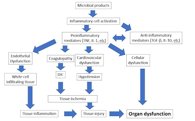
Sepsis Updated: 06/28/2022 © Jun Wang, MD, PhD   Definition Life threatening organ dysfunction caused by a dysregulated host response to infection General features Organ dysfunction identified by acute change of 2 or more points in total SOFA score or qSOFA Likely prolonged ICU stay Relatively high mortality rate Commonly associated with infections of injury or internal organs and perforation/rupture of abdominal/pelvic structures Most commonly caused by bacterial infections, but can be caused by virus or fungi Systemic Inflammatory Response Syndrome (SIRS) A term being abandoned since 2016 Still being used clinically Poor mortality prediction comparing with Sequential Organ Failure Assessment (SOFA) Clinical responses to either infectious or non-infectious causes Sequential Organ failure Assessment (SOFA) A systemic evaluation of organ function Performed in critically ill patients with suspected sepsis Systems involved o    Cardiovascular: Blood pressure o