Posts

Showing posts with the label renal mass

Pediatric Wilms tumor

Pediatric Wilms tumor Updated: 03/10/2021 © Jun Wang, MD, PhD   General features Most common pediatric renal tumor Majority diagnosed prior to age 6 years Usually sporadic May be associated with WAGR syndrome , Denys-Drash syndrome, Beckwith-Wiedemann syndrome, nephroblastomatosis Metastasis: commonly lymph nodes, lung, liver, etc Most patients survive with current multimodality therapy WAGR syndrome Complex of congenital developmental abnormalities including aniridia, genitourinary malformations, and mental retardation Deletion of the short arm of chromosome 11: WT1 , PAX6 High risk for Wilms tumor Denys-Drash syndrome Triad of congenital nephropathy, Wilms tumor and intersex disorders WT1 mutation Beckwith-Wiedemann syndrome Most common overgrowth syndrome in infancy Triad of congenital exomphalos, macroglossia, and gigantism Mental retardation common Abnormalities 11p15.5 ( WT-2 ) Complex pathogenesis Probably associated with IGF-2 action Wilms tumor: most common
Simple cyst

Simple cyst   Updated: 10/06/2020 © Jun Wang, MD, PhD General features Commonly incidental finding May present as mass Calcification or hemorrhage may occur Clinical features Commonly asymptomatic Hemorrhagic cysts: Acute flank pain Pathological findings Thin-walled cortical cysts Filled with clear yellow fluid Single layer of cuboidal, flattened or atrophic epithelium Diagnosis Radiologic studies: Sonography, CT, MRI Management Percutaneous aspiration Continuous negative-pressure catheter drainage Sclerotherapy Back to kidney masses Back to contents

Urothelial carcinoma of renal pelvis

Urothelial carcinoma of renal pelvis   Updated: 10/27/2022 © Jun Wang, MD, PhD General features Commonly multifocal, probably through drop metastases May coexist with lower urinary tract urothelial carcinoma More common in male, mean age 67 Likely non-invasive if low grade Risk factors Tobacco use Phenacetin Industrial carcinogen exposure (coal, asphalt, petrochemicals, tar) Thorium containing radiologic contrast material Balkan endemic nephropathy Clinical features Hematuria Flank pain Pathological features Exophytic mass Papillary architecture lined by atypical urothelial cells Nests, clusters and single neoplastic cells with urothelial differentiation Markers Positive for p63, uroplakin , CK7, CK20, PAX8 Genetic abnormality Microsatellite instability Complex karyotype if high grade Diagnosis Radiologic studies Ureteropyeloscopy with cystoscopy Biopsy Urine cytology with molecular technologies such as FISH (Urovysion): only for

Renal papillary adenoma

Renal papillary adenoma   Updated: 10/06/2020 © Jun Wang, MD, PhD General features AKA renal cortical adenoma, benign Commonly incidental finding Increasing numbers during life Probably a precursor for papillary renal cell carcinoma Clinical features Commonly asymptomatic Pathological findings Usually < 5 mm Well-circumscribed, unencapsulated tumor in cortex Dense papillary/tubular/tubulopapillary architecture Small cuboidal cells without significant atypia Psammoma bodies, xanthoma cells may present NO invasion Back to kidney masses Back to contents

Papillary type renal cell carcinoma

Papillary type renal cell carcinoma   Updated: 10/06/2020 © Jun Wang, MD, PhD General features Originated from proximal or distal convoluted tubule May be hereditary Commonly multifocal (80% of tumors), occasionally bilateral Two types Type I: better prognosis, MET mutation, simple epithelium Type II: aggressive, associated with hereditary leiomyomatosis and renal cell cancer syndrome, pseudostratified epithelium Prognosis better than clear cell carcinoma Pathological features Thick capsule Papillary or tubulopapillary tumor lined by simple epithelium with low nuclear grade (type I)   or pseudostratified epithelium with more pleomorphic nuclei (type II) Foamy macrophages and intracellular hemosiderin Genetic abnormality MET Frequently trisomy 7, 17 Back to renal cell carcinoma Back to kidney masses Back to contents

Medullary cystic kidney disease

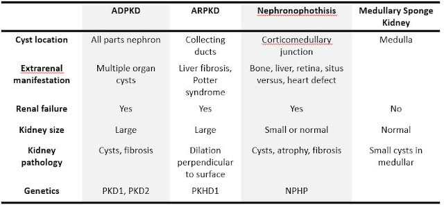
Medullary cystic kidney disease   Updated: 10/04/2018 © Jun Wang, MD, PhD General features Autosomal dominant AKA autosomal dominant interstitial kidney disease (ADIKD) or autosomal dominant tubulointerstitial kidney disease (ADTKD) No extrarenal involvement Three subtypes Uromodulin kidney disease (UKD) ADTKD DUE TO MUTATIONS IN THE REN GENE (ADTKD-REN) Mucin-1 kidney disease (MKD) Pathogenesis Loop of Henle deficiency due to uromodulin mutation (UKD) Accumulation of prerenin in tubules due to renin mutation (ADTKD-REN) Abnormal intracellular localization of mucin-1 in renal tubules (MKD) Clinical features Family history of kidney disease and gout Adult-onset progressive renal failure Early onset gout (teenage years) Pathological findings Similar to nephronophthisis Back to kidney masses Back to contents

Nephronophthisis

Image
Nephronophthisis   Updated: 09/30/2021 © Jun Wang, MD, PhD General features Heterogeneous group Autosomal recessive Most common genetic cause of pediatric end stage kidney disease Characterized by corticomedullary cysts, atrophy and interstitial fibrosis Diagnosis based on clinical findings and confirmed by genetic testing Key pathogenesis Cilia/basal body abnormality Abnormal renal tubular development and function Clinical presentations Infantile Most severe form Bilateral disease Median age for renal failure: 1 year of age Extrarenal presentations common: retinitis pigmentosa, hepatic fibrosis, skeletal / CNS malformations, situs inversus, etc Juvenile form Most common Polyuria/polydipsia due to cortical and tubulointerstitial damage Progresses to chronic renal failure in 5-10 years Anemia and growth retardation May be associated with retinitis pigmentosa Adolescent form : Similar to juvenile with later presentation (median age renal failur

Multicystic Renal Dysplasia

Multicystic Renal Dysplasia   Updated: 10/05/2020 © Jun Wang, MD, PhD General features AKA multicystic dysplastic kidney Congenital Most common pediatric cystic renal disease Usually sporadic but may be familial Bilateral involvement usually results in stillbirth or death within the first few days of life  Pathogenesis Abnormal induction of metanephric mesenchyme by ureteral bud Clinical features No significant clinical problem if unilateral Abdominal mass: Most common cause of abdominal mass in the newborns Commonly associated with ureteropelvic obstruction, ureteral agenesis, atresia or reflux May presents with urinary tract infection, voiding dysfunction, or hypertension Radiologic findings Sonographic findings: Multiple cysts Voiding cystourethrography: Vesicoureteral reflux Dimercaptosuccinic acid (DMSA) renal scanning: Loss of renal function, used if sonographic examination inconclusive Pathological findings Commonly unilateral with con

MiT translocation subgroup carcinomas

MiT translocation subgroup carcinomas   Updated: 10/07/2020 © Jun Wang, MD, PhD General features MiT: microphthalmia associated transcription factors Gene fusion involving TFE3 transcription factor on Xp11.2 or TFEB gene fusion due to t(6;11) Diagnosis requires FISH or PCR More common in women More common in pediatric and young adult population Aggressive clinical course Pathological features TFE3 rearrangement Papillary and nested growth pattern Eosinophilic and clear discohesive pseudostratified cells t(6:11) TFEB  Biphasic, small and large tumor cells Markers Positive: TFE3, TFEB May be positive: HMB45, melan A Genetic abnormality Translocation involving X11p: TFE3 transcription facto t(6:11): TFEB Diagnosis FISH or PCR Back to renal cell carcinoma Back to kidney masses Back to contents

Medullary Sponge Kidney

Image
Medullary Sponge Kidney   Updated: 09/30/2021 © Jun Wang, MD, PhD General features Sporadic Rarely progress to end stage renal disease due to complications (infections, nephrolithiasis) May be associated with hemihypertrophy of body, Marfan’s, Caroli’s and Ehlers-Danlos syndrome Etiology and pathophysiology Likely developmental abnormality Clinical features Usually asymptomatic with normal renal function Commonly incidental finding by image studies Adult onset of calcifications, stones, hematuria and infection May have hypercalcemia Pathological findings Small cystic dilation of medullary collecting ducts Normal cortex Diagnosis Intravenous pyelography or CT: R enal tubular ectasia Differential diagnosis Management Treatment of stones Treatment of flank pain Monitor renal function Back to kidney masses Back to contents

Hypoplasia of kidney

Hypoplasia of kidney   Updated: 10/05/2020 © Jun Wang, MD, PhD General features Congenital smaller kidney Unilateral or bilateral Reduced number of nephrons Hypertrophied nephron Often with dysplasia (hypodysplasia) Key pathogenesis Probably developmental defect of metanephric renal blastema Clinical features Oligohydramnios Presentations of renal impairment May develop end-stage renal disease Key morphological features Reduced number of nephrons Normal architecture Hypertrophic glomeruli/tubules with Thickened Bowman’s capsule Abnormal glomerular basement membrane Dysplasia common Diagnosis Image studies Back to kidney masses Back to contents

Horseshoe kidney

Horseshoe kidney Updated: 10/03/2018 © Jun Wang, MD, PhD General features Most common congenital kidney anomaly Most common fused at lower poles If complete fusion: pancake kidney (formless mass in the pelvis) May have anomalous superior vena cava May develop cancers, renal cell CA most common, followed by urothelial CA and sarcoma Risks for Wilms tumor and carcinoid Key pathogenesis Unclear Mechanical fusion: Touch and fusion Teratogenic: Abnormal migration of nephrogenic cells Clinical features Commonly asymptomatic Hydronephrosis common Symptoms associated with obstruction, stones, infection Urinary tract infection: Most common symptom in children Diagnosis Sonography Voiding cystourethrogram for vesicoureteral reflux Evaluations Renal function Back to kidney masses Back to contents

Ectopic kidney

Ectopic kidney Updated: 10/03/2018 © Jun Wang, MD, PhD General features Abnormal localization of kidney Unilateral or bilateral Most common sites: pelvic brim Key pathogenesis Cephalic migration arrest Clinical features Usually asymptomatic Risk of infections, trauma Back to kidney masses Back to contents

Collecting duct renal cell carcinoma

Collecting duct renal cell carcinoma   Updated: 05/05/2021 © Jun Wang, MD, PhD General features Aggressive Likely from distal collecting ducts More common in male, mean age 55 Commonly metastases at presentation to regional lymph nodes, bone, adrenal glands, lung, skin Pathological features Infiltrative tumor in medulla Irregular glands lined by high grade cells Numerous mitosis Marked fibrosis Back to renal cell carcinoma Back to kidney masses Back to contents

Clear cell type renal cell carcinoma

Clear cell type renal cell carcinoma   Updated: 10/06/2020 © Jun Wang, MD, PhD General features 70-80% of adult renal epithelial tumors Derived from proximal convoluted tubule Metastasis: commonly likely lung, lymph nodes, liver, bone, adrenal glands, etc, anytime, anywhere Risk factors Tobacco: Most significant risk factor Adult polycystic kidney disease Obesity, especially in women Hypertension Hereditary renal cell carcinoma without VHL Tuberous sclerosis von Hippel-Lindau syndrome Pathological features Well circumscribed (pushing borders) Orange/yellow (from lipid) Commonly with hemorrhage, necrosis and calcification Neoplastic cells with clear cytoplasm , arranged in nests with intervening blood vessels Markers Co-express cytokeratin and vimentin Genetic abnormality 3p- (3p25, VHL gene) Back to renal cell carcinoma Back to kidney masses Back to contents

von Hippel-Lindau syndrome

von Hippel-Lindau syndrome   Updated: 10/01/2021 © Jun Wang, MD, PhD General features Inherited disorder characterized by the formation of tumors and fluid-filled cysts in many different parts of the body Two types based on risk for pheochromocytoma Type 1. Low risk for pheochromocytoma only (1A) or low risk for both pheochromocytoma and renal cell carcinoma , (1B)  Type 2. High risk for pheochromocytoma Autosomal dominant inheritance Associated tumors appear at young adult age High risk of renal cell carcinoma , adrenal pheochromocytoma, pancreatic neuroendocrine tumor , retinal/CNS hemangioblastoma Clinical features Cysts in kidneys, pancreas, genital tract etc Hemangioblastoma Renal cell carcinoma Pheochromocytoma Genetic abnormality VHL: Encodes Von Hippel-Lindau disease tumor suppressor Alteration of both copies of the VHL gene needed for tumor and cyst formation Diagnosis Genetic testing: Detection of a germline mutation in the VHL gene Patients suspe

Oncocytoma

Oncocytoma Updated: 10/06/2020 © Jun Wang, MD, PhD General features Most common benign solid renal tumor Probably arising from intercalated cells More common in men May be associated with tuberous sclerosis complex (TSC) and Birt-Hogg-Dube syndrome if multiple and bilateral May coexist with renal cell carcinoma Clinical presentations Usually asymptomatic Rarely presents with hematuria and dysuria Flank pain if large Pathological findings Well circumscribed, unencapsulated, solid mass Stellate central scar Uniform round/polygonal cells Eosinophilic and granular cytoplasm and uniform small, round and central nuclei Tumor cells have abundant mitochondria Markers Scattered or negative CK 7 (positive for chromophobe renal cell carcinoma ) Negative: Hale’s colloidal iron stain (positive for chromophobe renal cell carcinoma ) Diagnosis Image studies Biopsy Management Excision Back to kidney masses Back to contents

Chromophobe type renal cell carcinoma

Chromophobe type renal cell carcinoma   Updated: 10/07/2020 © Jun Wang, MD, PhD General features Usually > 45, no gender preference Originated from intercalated cell of cortical collecting duct May be associated with Birt-Hogg-Dubé syndrome 8% multifocal, 3% bilateral Less aggressive comparing with clear cell type Birt-Hogg-Dubé syndrome Autosomal dominant with incomplete penetrance Germline mutations in BHD Skin lesions: fibrofolliculomas, trichodiscomas, acrochordons Lung: cysts, bullous emphysema, spontaneous pneumothorax Renal tumors: Oncocytoma, chromophobe type renal cell carcinoma, and hybrid oncocytic-chromophobe tumors with areas of clear cells Pathological features Well-circumscribed, tan brown Geographic necrosis Abundant slightly granular, pale eosinophiliccytoplasm Raisinoid nuclei and perinuclear halos Markers Diffusely positive for CK7 (Scattered to negative reactivity in oncocytoma ) Positive for Hale's colloidal iron stain (Ne

Renal cell carcinoma

Renal cell carcinoma   Updated: 05/05/2021 © Jun Wang, MD, PhD General features Most common type of kidney cancer in adults Usually > 60 years old, but younger if in familial clusters More common in male Rarely bilateral More common in people of Northern European ancestry (Scandinavians) and North Americans In the US, slightly more common in blacks Risk factors Tobacco: Most significant risk factor Obesity, especially in women Hypertension Longer usage of NSAID Long term renal dialysis, probably associated with acquired cystic disease Kidney transplantation Genetic disorders:   von Hippel-Lindau syndrome , hereditary papillary renal carcinoma , Birt-Hogg-Dube syndrome , and hereditary leiomyomatosis and renal cell cancer syndrome Clinical features Various, most asymptomatic until advanced stage Flank pain Hematuria Abdominal mass Vascular manifestations: scrotal varicocele, inferior vena cava involvements (edema, ascites, abnormal liver functions, et

Autosomal Recessive Polycystic Kidney Disease

Image
Autosomal Recessive (Childhood) Polycystic Kidney Disease   Updated: 10/01/2021 © Jun Wang, MD, PhD General features Many patients die shortly after birth Cause of death: Secondary respiratory insufficiency due to pulmonary hypoplasia Prognosis depends on kidney and liver involvements Key pathogenesis Probably abnormal centromere duplication and mitotic spindle assembly during cell division Developmental defects of collecting ducts Developmental defects of hepatobiliary ductal plate remodeling key clinical features Presentations associated with kidney and liver dysfunction Oligohydramnios Pulmonary hypoplasia: Respiratory distress Portal hypertension Potter syndrome Results of severe oligohydramnios Limb deformities: club feet, hip dislocation, etc Facial appearances: pseudoepicanthus, recessed chin, posteriorly rotated, flattened ears, and flattened nose Pulmonary hypoplasia Laboratory findings: High BUN/Creatinin Hyponatremia: Renal dysfunction