Posts

Showing posts from October, 2018

Nasopharyngeal carcinoma

Nasopharyngeal carcinoma   Updated: 10/13/2020 © Jun Wang, MD, PhD General features Arising from the epithelium of the nasopharynx Common in South Asia, North Africa, Middle East and the Arctic More common in men Rare in the states, but incident increasing in black teenagers Bimodal age distribution, a small peak in late childhood, and second peak around 50-59 Risk factors Associated with Epstein-Barr virus infection Other risk factors: Consumption of salt-preserved fish containing carcinogenic nitrosamines , family history, specific HLA class I genotypes, tobacco smoking , chronic respiratory tract conditions and low consumption of fresh fruits and vegetables Pathogenesis EBV genome products (LMP1, LMP2, etc) associated telomerase dysregulation, bcl2 overexpression, etc. Loss of function mutation of NF-kappa B inhibitors (CYLD, TRAF3, etc) Multiple chromosomal abnormalities p16 deletion LTBR (lymphotoxin-beta receptor) amplification Clinical presentati...

Vocal cord polyp

Vocal cord polyp   Updated: 10/13/2020 © Jun Wang, MD, PhD General features AKA laryngeal nodule or singer’s nodule Noninflammatory response to chronic irritation More common in heavy smokers or singers Almost never transforms to malignancy Etiology Chronic irritation including phonotrauma, allergies, smoking, etc Clinical presentations Generalized and persistent hoarseness Key morphological features Round growth with smooth surface on true vocal cords Fibrotic edematous core with reactive squamous epithelium Treatment Surgery Vocal rest Back to respiratory tract neoplasms Back to contents

Sinonasal papilloma

Sinonasal papilloma   Updated: 10/12/2020 © Jun Wang, MD, PhD General features AKA Schneiderian papilloma Benign Usually adults, 2/3 men Three subtypes Inverted (Most common): Risk for malignant transformation Fungiform (exophytic) : No known risk for malignant transformation Oncocytic : Risk for malignant transformation Clinical presentations Nasal obstruction, stuffiness or epistaxis Key pathogenesis Inverted: High and low risk HPV, EGFR mutation Fungiform : Low risk HPV Oncocytic : KRAS mutation HPV associated tumorigenesis Key morphological features Soft to moderately firm, with granular or finely clefted surface May have malignant foci Inverted papilloma : Endophytic growth , non keratinizing squamous, transitional or respiratory epithelium Fungiform papilloma : Exophytic growth , non keratinizing squamous, transitional or respiratory epithelium Oncocytic papilloma : Oncocytic epithelium, intraepithelial mucin-filled cysts and microabscesses ...

Olfactory neuroblastoma

Olfactory neuroblastoma   Updated: 10/12/2020 © Jun Wang, MD, PhD General features AKA esthesioneuroblastoma Rare, malignant neuroectodermal tumor Probably arise from olfactory membrane or olfactory placode NOT related to neuroblastomas elsewhere in body Mean age 53 years, range 3-79 years Slightly more common in male Late recurrence common Metastasis: Cervical lymph nodes, lungs Clinical presentations Usually upper nasal vault; rarely in nasopharynx, maxillary or ethmoid sinus Nasal obstruction, epistaxis Neurologic presentations: headache, nausea, diplopia, etc Rarely paraneoplastic syndromes : Hypercalcemia, ectopic adrenocorticotropic hormone syndrome, etc Presentations associated with local invasion Radiologic findings Homogeneous soft tissue mass with uniform and moderate contrast enhancement Key morphological features Nests or sheets of uniform small cells with round nuclei Markers Positive for cytokeratin, neuron-specific enolase, s...

NUT carcinoma

NUT carcinoma   Updated: 10/12/2020 © Jun Wang, MD, PhD General features Formerly known as NUT midline carcinoma Aggressive type of poorly differentiated squamous cell carcinoma Named after molecular abnormality involving NUT (nuclear protein in testis) gene Commonly midline location, including head and neck, mediastinum Metastasis common Clinical presentations Mass related symptoms Key morphological features I nfiltrating trabeculae of undifferentiatedcells   Sudden squamous differentiation Desmoplastic stroma May have foci of squamous differentiation Marker Nuclear expression of NUT Genetic abnormality Nuclear protein of the testis (NUT) at 15q14 Diagnosis Morphology Molecular tests (fluorescence in situ hybridization) for translocations involving NUT Treatment No effective treatment Back to respiratory tract neoplasms Back to contents

Nasopharyngeal angiofibroma

Nasopharyngeal angiofibroma   Updated: 10/12/2020 © Jun Wang, MD, PhD General features Rare, benign Adolescent and young adult males Locally aggressive Likely recur Clinical presentations Nasal obstruction, epistaxis Commonly posterolateral wall of roof of nose Key morphological features Polypoid mass Well circumscribed Stellate and staghorn blood vessels Fibrotic background Markers Commonly express androgen receptor Variable estrogen or progesterone receptors Treatment Surgery Back to respiratory tract neoplasms Back to contents

Squamous papilloma and papillomatosis

Image
Upper respiratory squamous papilloma and papillomatosis   Updated: 10/12/2020  © Jun Wang, MD, PhD General features Warty outgrowths of surface epithelium More common in men Commonly multiple in children, but single in adult Likely recur Etiology Squamous neoplasm associated with HPV, especially HPV 6 and 11 Clinical presentations Airway obstruction or voice change Key pathogenesis HPV associated tumorigenesis Key morphological features (Image credit: Nephron [CC BY-SA 3.0 (https://creativecommons.org/licenses/by-sa/3.0)]) Fibrovascular cores Well-differentiated squamous epithelium with koilocytotic change Dysplasia may be seen in adults Treatment Goals of therapy: Relieve airway obstruction, improve voice quality, and facilitate remission Surgery Antiviral agent: Cidofovir Back to respiratory tract neoplasms Back to contents

Laryngeal squamous cell carcinoma

Laryngeal squamous cell carcinoma Updated: 10/12/2021 © Jun Wang, MD, PhD General features Majority of laryngeal cancers Predominantly male; usually ages 40+ Risk factors Smoking , enhanced by heavy alcohol consumption HPV Clinical presentations Dysphonia/aphonia Dysphagia Blood-tinged sputum, etc   Metastases: Regional lymph nodes and lungs Direct extension to surrounding structures: Thyroid gland and jugular vein Key pathogenesis HPV associated tumorigenesis: E6 and E7 Key morphological features Mass, possibly ulcerated Irregular nests, cords of atypical squamous cells (Large nuclei, irregular nuclear shape and size, irregular chromatin pattern) Squamous differentiation (intercellular bridges, squamous pearls) Treatment Goals of therapy: Remove the tumor and prevent recurrence while maintaining laryngeal function Surgery, radiation, chemotherapy Poor prognostic factors High initial staging Tumor grade Vascular invasion Negative ...

Branchial pouch/cleft anomalies

Branchial pouch/cleft anomalies   Updated: 10/13/2020 © Jun Wang, MD, PhD General features Congenital lesions Usually 20-40 years old Cysts most common May be bilateral Clinical presentations May be cyst, sinus, fistula or cartilage in anterolateral neck   Key pathogenesis Incomplete obliteration of the branchial apparatus Key morphological features Cysts derived from branchial cleft have squamous epithelium Cysts derived from branchial pouch have respiratory epithelium, with or without squamous metaplasia Branchial cyst Congenital epithelial cysts Lateral neck Failure of obliteration of the second branchial cleft Solitary, painless mass in the neck of a child or a young adult Intermittent swelling and tenderness during upper respiratory tract infection Fistula after inflammation Cyst with stratified squamous or ciliated columnar epithelium and nonspecific inflammation Treatment: Surgery, antibiotics if infected Thyroglossal duct c...

Solitary fibrous tumor

Solitary fibrous tumor   Updated: 02/17/2023 © Jun Wang, MD, PhD General features Pleural based, but usually NO pleural effusion Most likely from submesothelial mesenchyme Not associated with asbestos May occur at other locations May cause death due to extensive intrathoracic growth or malignant type Aggressive if malignant Key clinical features May be asymptomatic Nonspecific pulmonary symptoms including cough, shortness of breath, or chest pain, etc Associated with pulmonary osteoarthropathy, digital clubbing and hypoglycemia, which regress after tumor resection Key morphological features Firm well circumscribed, pleural based mass Markedly collagenous stroma with irregularly distributed thick walled " staghorn " vessels and bland spindle cells Malignant solitary fibrous tumor: marked pleomorphism , mitosis , necrosis Markers Positive: CD34, CD99, STAT 6, vimentin Negative: Cytokeratin Genetic abnormality Fusion of NAB2-STAT6  Treatmen...

Mesothelioma

Mesothelioma   Updated: 10/15/2020 © Jun Wang, MD, PhD General features Arises from mesothelial lining of pleura, peritoneum, pericardium and tunica vaginalis Pleura most common location More common in men, 50-80 years old May occur in younger population with equal sexual distribution Poor prognosis, commonly recur Key risk factors Asbestos exposure Peak age 35-45 years after asbestos exposure Other risk factors: Radiation, Erionite, SV40 virus Key pathogenesis Direct damage of asbestos: Toxicity, ROS production, DNA damage Immune suppression Key clinical features Most common presentations: Dyspnea and nonpleuritic chest wall pain Other presentations: Chest discomfort, pleuritic pain, sweat, etc Key morphological features Likely multifocal Epithelioid : tubulopapillary , glandular , solid , etc Sarcomatoid: Malignant spindle cell proliferation   Mixed epithelioid and sarcomatoid components Genetic abnormalities P16 INK4 a /p14 ARF N...

Small cell carcinoma of lungs

Small cell carcinoma of lungs   Updated: 10/15/2020 © Jun Wang, MD, PhD General features Also called oat cell carcinoma Poorly differentiated neuroendocrine tumor Derived from primitive cells of basal bronchial epithelium More common in men, median age 60 Strong association with tobacco smoking Most common lung cancer associated with uranium mining , probably associated with radon Smoking cessation associated with improved survival Key clinical features Nonspecific presentations similar to other lung cancer: cough, dyspnea, etc Associated paraneoplastic syndromes due to production of ACTH, ADH, calcitonin, etc Lambert-Eaton myasthenic syndrome Presynaptic disorder of neuromuscular transmission Autoimmune process against voltage-gated calcium channels (VGCCs) Occasionally antibodies against acetylcholine receptor , but usually low titers (High titer seen in myasthenia gravis) Impaired release of acetylcholine Presentations: Proximal muscle weakness ...

Squamous cell carcinoma of lungs

Squamous cell carcinoma of lungs   Updated: 10/15/2020 © Jun Wang, MD, PhD General features Arising from squamous epithelial cells (likely after metaplasia and dysplasia) Used to be most common type of lung cancer in Western countries Rates declining due to reduction in tobacco use Usually men, closely correlated with smoking history Key risk factor Smoking Clinical presentations Bronchial obstruction (pneumonitis, atelectasis) Associated paraneoplastic syndrome: Hypercalcemia due to cancer production of parathyroid hormone-related peptide Commonly seen molecular abnormalities p53 Amplification: PI3KCA, SOX2, FGFR, MET Mutation: PTEN Key morphological features Mass, usually centrally located Irregular nests, cords of atypical cells (Large nuclei, irregular nuclear shape and size, irregular chromatin pattern) Squamous differentiation ( intercellular bridges, squamous pearls ) Markers CK5/6, p63 Treatment Surgery Radiation Chemotherap...

Large cell carcinoma of lungs

Large cell carcinoma of lungs   Updated: 11/10/2022 © Jun Wang, MD, PhD General features Undifferentiated malignant epithelial tumor composed of large, atypical cells Associated with smoking More common in men, age 50 and up Diagnosis of exclusion only on resected tumor Key morphological features Mass, usually peripheral location Frequently involves thoracic walls Usually solid nests Markedly atypical and anaplastic cells Markers Positive: Cytokeratin May be negative for squamous and adenocarcinoma markers, as well as neuroendocrine markers, including p63, TTF1, chromogranin, CD56, etc Treatment Surgery Radiation Chemotherapy     Back to malignant lung tumors Back to respiratory tract neoplasms Back to contents

Carcinoid of lungs

Well-differentiated neuroendocrine tumor of lung   Updated: 10/15/2020 © Jun Wang, MD, PhD General features Well-differentiated neuroendocrine neoplasm Locally invasive, rarely metastasizes more common in female, age <60 May have MEN1 mutation NOT related to smoking Key clinical features May be asymptomatic Symptoms of bronchial obstruction Presentations associated with hormone production Key morphological features Well defined border No necrosis Nests or trabeculae of cells with lightly eosinophilic cytoplasm, round to oval nuclei Salt and pepper chromatin pattern Markers Positive: Cytokeratin Positive for neuroendocrine markers: CD56, neuron specific enolase, chromogranin, synaptophysin Negative: CD45 and other lymphocytic markers Treatment Excision Back to malignant lung tumors Back to respiratory tract neoplasms Back to contents

Malignant neoplasms of lungs

Image
Malignant neoplasms of lung   Updated: 07/11/2022 © Jun Wang, MD, PhD General features Leading cause of cancer death in the United States Cessation of smoking improves prognosis Metastasis commonly to adrenal glands , brain, contralateral lung, bone, liver, etc Risk factors Cigarette smoking : Most common risk factor; quantity of cigarette smoking summarized by the number of packs of cigarettes smoked per day multiplied by the number of years smoked Secondhand smoke Radiation at home: Radon Occupational and environmental factors: wood/coal fuels , asbestos , arsenic, radiation, dusts and fumes from nickel, chromium, etc Family and genetic factors Clinical presentations Coughing Hemoptysis Chest pain Others: Pneumonia, pleural effusion, etc Superior vena cava syndrome Medical emergency and most often manifests in patients with a malignant disease process within the thorax Due to obstruction of blood flow through the superior vena cava May cause obs...

Adenocarcinoma of lungs

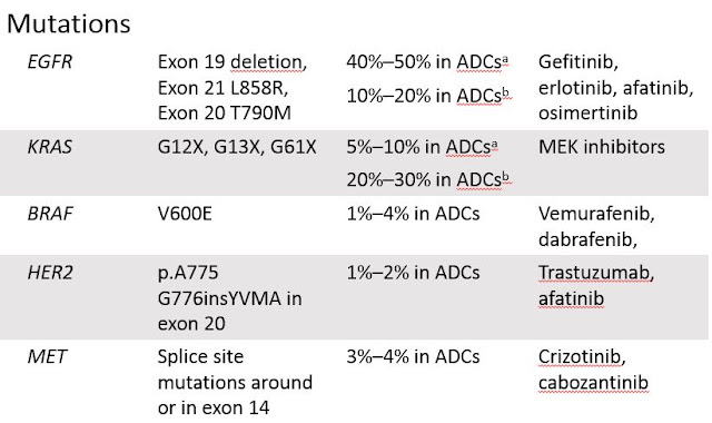
Adenocarcinoma of lungs   Updated: 10/15/2020 © Jun Wang, MD, PhD General features Arising from bronchi, bronchioles and alveolar cells More common in female Most common type of lung cancer in nonsmokers males Clinical presentations Bronchial obstruction (pneumonitis, atelectasis) Associated paraneoplastic syndrome Clubbing Hypertrophic pulmonary osteoarthropathy  Clubbing Spoon-shaped nails Inflammation, swelling and pain in the hand, fingers, knees or ankles Trousseau syndrome of hypercoagulability Commonly seen molecular abnormalities p53 Amplification: MET, Her2 Mutation: EGFR, KRAS, PTEN, LKB Fusion: EML4-ALK KRAS mutation and MET amplification associated with poor prognosis and acquired EGFR inhibitor resistance Key morphological features Glandular differentiation (single layer of cuboidal to columnar cells, mucin production, luminal spaces) With or without mucin production  Irregular glands if differentiated Papillary ...

Neoplasms of respiratory tract

Neoplasms of respiratory tract   Updated: 11/14/2022 © Jun Wang, MD, PhD Anatomy Nasal cavity Nasopharynx Oropharynx Laryngopharynx Larynx Trachea and bronchi Lungs Histology Squamous mucosa Respiratory mucosa May have abundant lymphoid tissue in pharynx Type I and Type II pneumocytes Key risk factors Human papillomavirus: Squamous cell carcinoma, squamous cell papilloma Epstein Barr virus: Nasopharyngeal carcinoma Smoking Tumors of nasal cavity and nasopharynx Olfactory neuroblastoma Nasopharyngeal angiofibroma Sinonasal papilloma NUT midline carcinoma Nasopharyngeal carcinoma Tumors or tumor like lesions of larynx Vocal cord polyp Squamous papilloma Squamous cell carcinoma Branchial pouch/cleft anomalies Branchial cyst Thyroglossal duct cyst Neoplasms of lung Squamous cell carcinoma Adenocarcinoma Large cell carcinoma Small cell carcinoma Carcinoid Lymphangioleiomyomatosis Neoplasms of pleura Mesotheli...

Pediatric Wilms tumor

Pediatric Wilms tumor Updated: 03/10/2021 © Jun Wang, MD, PhD   General features Most common pediatric renal tumor Majority diagnosed prior to age 6 years Usually sporadic May be associated with WAGR syndrome , Denys-Drash syndrome, Beckwith-Wiedemann syndrome, nephroblastomatosis Metastasis: commonly lymph nodes, lung, liver, etc Most patients survive with current multimodality therapy WAGR syndrome Complex of congenital developmental abnormalities including aniridia, genitourinary malformations, and mental retardation Deletion of the short arm of chromosome 11: WT1 , PAX6 High risk for Wilms tumor Denys-Drash syndrome Triad of congenital nephropathy, Wilms tumor and intersex disorders WT1 mutation Beckwith-Wiedemann syndrome Most common overgrowth syndrome in infancy Triad of congenital exomphalos, macroglossia, and gigantism Mental retardation common Abnormalities 11p15.5 ( WT-2 ) Complex pathogenesis Probably associated with IGF-2 action Wilms tumor: most co...

Simple cyst

Simple cyst   Updated: 10/06/2020 © Jun Wang, MD, PhD General features Commonly incidental finding May present as mass Calcification or hemorrhage may occur Clinical features Commonly asymptomatic Hemorrhagic cysts: Acute flank pain Pathological findings Thin-walled cortical cysts Filled with clear yellow fluid Single layer of cuboidal, flattened or atrophic epithelium Diagnosis Radiologic studies: Sonography, CT, MRI Management Percutaneous aspiration Continuous negative-pressure catheter drainage Sclerotherapy Back to kidney masses Back to contents

Urothelial carcinoma of renal pelvis

Urothelial carcinoma of renal pelvis   Updated: 10/27/2022 © Jun Wang, MD, PhD General features Commonly multifocal, probably through drop metastases May coexist with lower urinary tract urothelial carcinoma More common in male, mean age 67 Likely non-invasive if low grade Risk factors Tobacco use Phenacetin Industrial carcinogen exposure (coal, asphalt, petrochemicals, tar) Thorium containing radiologic contrast material Balkan endemic nephropathy Clinical features Hematuria Flank pain Pathological features Exophytic mass Papillary architecture lined by atypical urothelial cells Nests, clusters and single neoplastic cells with urothelial differentiation Markers Positive for p63, uroplakin , CK7, CK20, PAX8 Genetic abnormality Microsatellite instability Complex karyotype if high grade Diagnosis Radiologic studies Ureteropyeloscopy with cystoscopy Biopsy Urine cytology with molecular technologies such as FISH (Urovysion): only ...

Renal papillary adenoma

Renal papillary adenoma   Updated: 10/06/2020 © Jun Wang, MD, PhD General features AKA renal cortical adenoma, benign Commonly incidental finding Increasing numbers during life Probably a precursor for papillary renal cell carcinoma Clinical features Commonly asymptomatic Pathological findings Usually < 5 mm Well-circumscribed, unencapsulated tumor in cortex Dense papillary/tubular/tubulopapillary architecture Small cuboidal cells without significant atypia Psammoma bodies, xanthoma cells may present NO invasion Back to kidney masses Back to contents

Papillary type renal cell carcinoma

Papillary type renal cell carcinoma   Updated: 10/06/2020 © Jun Wang, MD, PhD General features Originated from proximal or distal convoluted tubule May be hereditary Commonly multifocal (80% of tumors), occasionally bilateral Two types Type I: better prognosis, MET mutation, simple epithelium Type II: aggressive, associated with hereditary leiomyomatosis and renal cell cancer syndrome, pseudostratified epithelium Prognosis better than clear cell carcinoma Pathological features Thick capsule Papillary or tubulopapillary tumor lined by simple epithelium with low nuclear grade (type I)   or pseudostratified epithelium with more pleomorphic nuclei (type II) Foamy macrophages and intracellular hemosiderin Genetic abnormality MET Frequently trisomy 7, 17 Back to renal cell carcinoma Back to kidney masses Back to contents