Practice question answers lower urinary tract pathology
Practice question answers
Lower urinary tract pathology
© Jun Wang, MD, PhD
1. C. Hematuria may
be caused by various disorders. Outpouching of ureter without atypical cells are
most consistent with diverticula.
Urothelial
carcinoma of ureter usually presents with mass (filling defects,
obstruction, etc) during image studies. In addition, all urothelial neoplasms
likely have atypical cells identified in urine, same as bladder
cancers.
2. E. The patients
presents with symptoms of bilateral urinary obstruction. Image studies reveal a
pelvic mass. Microscopic finding of fibrous tissue without atypia is most
compatible with a benign fibrotic process, i.e. sclerosing
retroperitoneal fibrosis, a condition of IgG4 associated diseases. The
plasma cells usually contains IgG4. Diffuse
large B cell lymphoma is characterized by sheets of markedly atypical cells
expressing CD20, not scattered lymphocytes and plasma cells. Fibrosarcoma
is characterized by hypercellular spindle cell growth with herringbone pattern,
and cytological atypia. Hodgkin
lymphoma has Reed Sternberg cells in a background of inflammatory cells. Plasmacytoma
is characterized by sheets of atypical plasma cells.
3. C. Sclerosing
retroperitoneal fibrosis, a condition of IgG4 associated diseases. The plasma cells usually contains
IgG4. Hypercalcemia can be seen in primary
or tertiary
hyperparathyroidism and tumor associated bone destruction. Elevated
serum CD30 can be seen in a few lymphomas, including Hodgkin
lymphoma. Elevated kappa light chain and monoclonal IgG can be seen in
plasma neoplasms, including plasmacytoma,
multiple
myeloma, monoclonal
gammopathy of undetermined significance and lymphoplasmacytic
lymphoma.
4. D. Small benign intramucosal
cystic lesion is most likely ureteritis
cystica, a condition associated with recurrent UT infection. It is seen as
filling defects protruding into lumen during urography exam. Diabetic
nephropathy is
associated with increased glomerular extracellular matrix, resulted from
hyperglycemia, and presents with proteinuria. It does not have ureter manifestations.
All urothelial neoplasms likely have atypical cells identified in urine,
same as bladder
cancers. Ureter diverticula
is characterized by outpouching of ureter without atypical cells.
5. A. Polypoid ureter
mass with fibrovascular core and benign urothelial lining is most compatible
with fibroepithelial
polyp. Low
grade papillary urothelial carcinoma have papillary growth with urothelial cytological
atypia. Ureter diverticula
is characterized by outpouching of ureter without atypical cells. Ureteritis
cystica is small benign intramucosal cystic lesion seen as filling defects protruding
into lumen during urography exam. Urothelial
carcinoma in situ is a flat lesion with marked cytological atypia.
6. B. Glandular
structure in bladder lamina propria is suspicious for adenocarcinoma. However, adenocarcinoma
of bladder, as adenocarcinoma in any other sites, has irregular gland and
cytological atypia. When both are absent, it is likely cystitis
glandularis. Low
grade papillary urothelial carcinoma and urothelial
papilloma have papillary growth, with or without urothelial cytological
atypia. Urothelial
carcinoma in situ is a flat lesion with marked cytological atypia.
7. C. Presentation
of bladder inflammation, with normal laboratory, cystoscopy and image studies
is most likely interstitial
cystitis. Acute
cystitis has evidence of bacterial infection, such as pyuria, positive
microbiology studies, etc. Cystitis
glandularis usually has raised lesions, due to clustered glandular
structures in lamina propria. Papillary
urothelial carcinoma and urothelial
papilloma have papillary growth, with or without urothelial cytological
atypia.
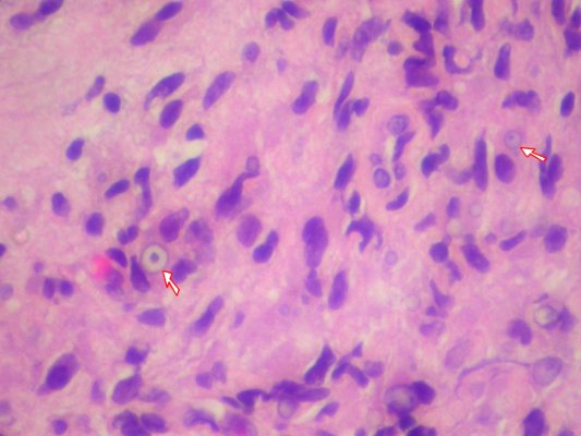
8. C. Aggregates of
macrophages with the presence of Michaelis-Gutmann
bodies (iron containing, cytoplasmic laminated mineralized concretions) is
diagnostic for malakoplakia.
Papillary
urothelial carcinoma and urothelial
papilloma have papillary growth, with or without urothelial cytological
atypia. Urothelial
carcinoma in situ is a flat lesion with marked cytological atypia.
9. E. Bladder papillary
growth with a fibrovascular core and a few layers of normal appearing urothelial
covering is urothelial
papilloma. Papillary
urothelial carcinoma has urothelial cytological atypia. Malakoplakia
has aggregates of macrophages with the presence of Michaelis-Gutmann
bodies. Polypoid
cystitis is polypoid growth associated with chronic bladder inflammation or
injury, such as catheterization. Urothelial
carcinoma in situ is a flat lesion with marked cytological atypia.
10. B. Urothelial
papilloma and papillary
urothelial carcinoma may have FGFR3 mutation, especially low grade cases.
EGFR, p53 and Rb are more commonly associated with high grade urothelial cancers,
including urothelial
carcinoma in situ. Racemase overexpression is seen in prostate
adenocarcinoma.
11. B. Papillary growth
with increased epithelial layers and mildly atypical urothelial nuclei, but no evidence of high grade nuclear features (marked variation in size, shape, and darkness) is most likely low
grade papillary urothelial carcinoma. High
grade papillary urothelial carcinoma has markedly cytological atypia. Squamous
cell carcinoma has squamous differentiation, such as intercellular
bridges and squamous pearls. Urothelial
carcinoma in situ is a flat lesion with marked cytological atypia. Urothelial
papilloma have no urothelial cytological atypia.
12. A. See
discussion of question 11.
13. D. See
discussion of question 11.
14. E. One of the
most important character for urothelial
carcinoma is multifocality, along the urinary tract, from renal pelvis to
bladder/urethra.
15. B. Presence of
cords of atypical epithelial cells in lamina propria/muscular layer is always a
sign of invasion. This is a case with mildly atypical urothelial cells invading
into bladder wall, consistent with low grade invasive
urothelial carcinoma. High
grade papillary urothelial carcinoma has markedly cytological atypia, and
is usually refer to non-invasive form. Normal Brunn nests have regular border
and no cytological atypia. Squamous
cell carcinoma has squamous differentiation, such as intercellular
bridges and squamous pearls. Urothelial
carcinoma in situ is a flat lesion with marked cytological atypia. Urothelial
papilloma have no urothelial cytological atypia.
16. D. Squamous cell
carcinoma of skin rarely metastasize, especially nowadays. Also see discussion of
question 15.
17. D. Schistosoma
haematobium infection is a leading cause of bladder
squamous cell carcinoma in patients from Sudan and Egypt. UV light is the leading
cause of skin cancers.
18. A. Irregular
glands lined by atypical cells are most compatible with adenocarcinoma,
regardless of the site. This case is a bladder adenocarcinoma.
Cystitis
glandularis has glands with smooth contours, instead of irregular glands
with features of invasion. Papillary
urothelial carcinoma, urothelial
carcinoma in situ and urothelial
papilloma do not have glandular differentiation.
19. B. Replacement
of urothelium by normal appearing intestinal type is consistent with intestinal
metaplasia, a risk factor for bladder adenocarcinoma. Adenocarcinoma
of bladder, as adenocarcinoma in any other sites, has irregular gland and
cytological atypia. Interstitial
cystitis usually does not have epithelial changes. Malakoplakia
has aggregates of macrophages with the presence of Michaelis-Gutmann
bodies.
20. A. Polypoid growth
of urethra with features of inflammation and granulation tissue is most
consistent with caruncle.
Condyloma
has papillary growth and koilocytic
changes. Herpes
has multinucleated cells with margining of chromatin and nuclear molding. Papillary
urothelial carcinoma and urothelial
papilloma have papillary growth, with or without urothelial cytological
atypia.
Back to contents
Comments
Post a Comment