Posts

Showing posts with the label lymphoid

Mycosis fungoides

Mycosis fungoides   Updated: 03/17/2021 © Jun Wang, MD, PhD General features Most common type of cutaneous T cell lymphoma More commonly seen in adults/elderly, men, African Unknown etiology Clonal CD4+ T cells Diagnosis based on clinical presentations, biopsy, molecular testing and immunopathologic profiling  Clinical presentations Four stages: Patch, plaque, tumoral and Sézary syndrome Patch stage: Pruritic erythematous macules or patches with telangiectasia and atrophy, may disappear spontaneously Plaque stage: Pruritic thichened plaques , may resemble psoriasis Tumoral stage: Tumor formation , either from plaques or de novo, may ulcerate Sézary syndrome Commonly erythroderma (80% of total body surface), may be scaly Lymphadenopathy Sézary cells in skin, lymph nodes and peripheral blood Usually do not evolve from patches, plaques or tumors May have marrow involvement Key morphological features Patch stage: Psoriasiform changes, but intraepidermal clonal
Adult T Cell leukemia/lymphoma

Adult T Cell leukemia/lymphoma Updated: 0 7/16/2019 © Jun Wang, MD, PhD General features Aggressive Endemic with Human T Cell Leukemia Virus Type I (HTLV-I) Clinical presentations Wide spread lymph node and peripheral blood involvement Key pathogenesis Human T cell leukemia virus Viral protein (Tax) activates NF- k B Key Laboratory findings Hypercalcemia due to increased osteoclastic activity Key morphological features Cloverleaf/flower cells : Neoplastic T cells with agranular basophilic cytoplasm, multilobated nuclei, in the appearance of clover leaf Markers Positive: CD3, CD4 Negative: CD30, ALK Back to lymphoid neoplasms Back to contents

Peripheral T-Cell Lymphoma, Unspecified

Peripheral T-Cell Lymphoma, Unspecified Updated: 08/24/2020 © Jun Wang, MD, PhD General features Heterogeneous group Mature T cell neoplasms Most common nodal T cell lymphoma Highly aggressive Usually high stage with skin and lung involvement Clinical presentations Nodal or extranodal Key morphological features Pleomorphic mixture of variably sized malignant T cells Markers Frequently loss of T-cell markers, such as CD5, CD7 Genetic abnormalities Clonal T-cell receptor rearrangement Back to lymphoid neoplasms Back to contents

Extranodal NK/T cell lymphoma, nasal type

Extranodal NK/T cell lymphoma, nasal type Updated: 07/16/2019 © Jun Wang, MD, PhD General features Rare, extranodal lymphoma Highly aggressive Express CD56 Clinical presentations Most common site: Upper aerodigestive tract Rapidly growing mass Vascular damage and destruction Prominent necrosis Key pathogenesis EB Virus Back to lymphoid neoplasms Back to contents

Large granular lymphocytic leukemia

Large granular lymphocytic leukemia Updated: 08/24/2020 © Jun Wang, MD, PhD General features Heterogeneous group Persistent increased peripheral blood large granular lymphocytes (LGL) May coexist with other hematopoietic neoplasms Clinical presentations Associated with autoimmune disorders, most common rheumatoid arthritis Key pathogenesis Unclear Key Laboratory findings Lymphocytosis Often severe neutropenia with or without anemia Aplastic anemia, thrombocytopenia, splenomegaly, etc Key morphological features Large lymphocytes with azurophilic granules Either T cell or NK cell Genetic abnormalities Activating mutation of STAT3 Back to lymphoid neoplasms Back to contents

Hodgkin lymphoma

Image
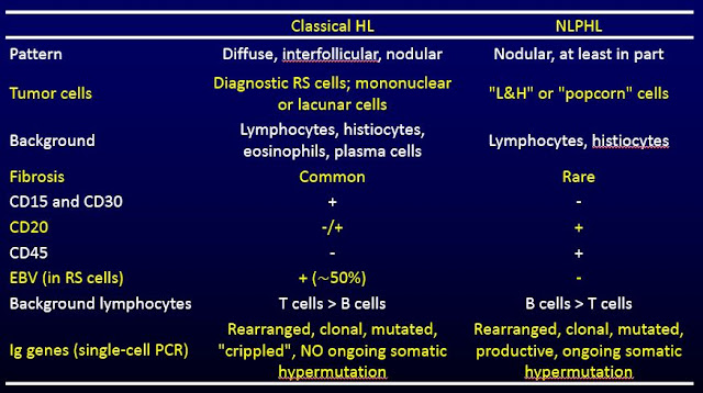
Hodgkin lymphoma   Updated: 07/03/2023 © Jun Wang, MD, PhD General features Usually nodal Likely B cell origin   Two large groups: classical or nodular lymphocyte predominant   Classical Hodgkin lymphoma Most common Hodgkin lymphoma Most likely B cell origin More common in HIV + population or history of infectious mononucleosis Sub classification of classical Hodgkin lymphoma based on background morphology Nodular lymphocyte predominant Hodgkin lymphoma Uncommon Probably follicular B cell origin More common in men Clinical presentations Lymphadenopathy B-type symptoms (fever, drenching night sweats, weight loss) Other organ involvement: Spleen, liver, lung, bone marrow, etc Classical Hodgkin lymphoma Bimodal age distribution in developed country: 15-35, > 54 Usually lymph nodes: Most common cervical, can be seen in mediastinal, axillary and paraaotic lymph nodes Rarely extranodal Nodular lymphocyte predominant Hodgkin lymphoma Indolent

Primary Effusion Lymphoma

Primary Effusion Lymphoma Updated: 08/24/2020 © Jun Wang, MD, PhD General features A type of Diffuse large B cell lymphoma  Also called body cavity large B cell lymphoma Advanced HIV infection High grade Aggressive Poor prognosis Clinical presentations Effusions Usually no identifiable tumor mass Key pathogenesis Human herpes virus 8 HIV EB virus Key Laboratory findings High grade tumor cells in effusions, pleural, pericardial or peritoneal Key morphological features of tumor cells Large pleomorphic lymphocytes Markers Positive: B cell markers including CD19, CD2, EBER Back to lymphoid neoplasms Back to contents

Diffuse Large B Cell Lymphoma

Diffuse Large B Cell Lymphoma   Updated: 08/24/2020 © Jun Wang, MD, PhD General features Most common non-Hodgkin lymphoma High grade Fast growing, aggressive Either de novo or transformed from other low grade lymphoma Most common testicle tumor in patient older than 65 WHO has subclassifications Clinical presentations Rapid growing mass, nodal or extranodal Systemic "B" symptoms: Fever, weight loss, drenching night sweats Key pathogenesis Bcl 6 associated NF-kB pathway Key morphological features of tumor cells Diffuse infiltration Sheets of tumor cells Pleomorphic large tumor cells Irregular size, shape, chromatin pattern, prominent nucleoli Markers Positive: B cell markers CD19, CD20 Negative: T cell markers CD3, CD4, CD8 Negative: Epithelial markers: cytokeratin, etc High proliferative index per ki67, but less than 100% Genetic abnormalities Various Translocations involving bcl2 or bcl 6 Back to lymphoid neoplasms Back

Lymphoplasmacytic lymphoma

Lymphoplasmacytic lymphoma Updated: 08/24/2020 © Jun Wang, MD, PhD General features B cell disorder with features of both low grade lymphoma and myeloma Older patients Frequently involving bone marrow, lymph node, spleen, liver Rarely lytic bone lesion, amyloidosis , renal impairment Autoimmune hemolysis may occur May transform into Diffuse large B celllymphoma Clinical presentations Bone marrow abnormality: Anemia, thrombocytopenia, neutropenia Non specific: Lymphoadenopathy, hepatosplenomegaly Hyperviscosity syndrome May be asymptomatic Key pathogenesis MYD88 mutation resulted in abnormal function of Toll-like receptor and Il-1 receptor signaling pathway Key Laboratory findings Waldenstrom macroglobulinemia Serum IgM monoclonal gammopathy, M spike Monoclonal Ig light chains ( Bence-Jones protein ) in urine Key morphological features Small lymphocyte Abnormal plasma cells similar to plasmacytoma Markers Positive: CD19, CD20, CD138, ligh

Multiple myeloma

Multiple myeloma Updated: 08/24/2020 © Jun Wang, MD, PhD General features Plasma cell neoplasm Clinical variants Asymptomatic (smoldering) myeloma : NO related organ or tissue impairment Non-secretory myeloma : NO M-protein by electrophoresis or immunofixation ; clinical features similar to secretory myeloma except for low incidence of renal insufficiency and hypercalcemia Plasma cell leukemia : Clonal plasma cells > 20% of the leukocyte differential count; aggressive disease Clinical presentations Widespread lytic bone lesions , bone pain, pathological fracture Renal insufficiency: caused by toxicity of light chains to renal epithelium Amyloidosis Hyperviscosity syndrome Increased risk of infection, most common cause of death Key pathogenesis DKK1 and FRP2: Inhibit Wnt, blocking bone formation Elevated RANKL, promotes osteoclasts activity, similar to giant cell tumor of bone Suppressed OPG Key Laboratory findings Monoclonal g

Monoclonal gammopathy of undetermined significance (MGUS)

Monoclonal gammopathy of undetermined significance (MGUS) Updated: 08/24/2020 © Jun Wang, MD, PhD General features Monoclonal immunoglobulin in serum or urine without clinical presentations of multiple myeloma Most common plasma cell dyscrasia May transform into multiple myeloma , amyloidosis , Waldenström macroglobulinemia or other lymphoma Follow up with serum paraprotein , but no specific treatment Clinical presentations Usually incidental finding of paraprotein , most common IgG May presents with paraproteinemic neuropathy , especially IgM No organ dysfunction as seen in multiple myeloma Key Laboratory findings Less than 10% clonal plasma cells in bone marrow   < 3 g/dl serum immunoglobulin Key morphological features Abnormal plasma cells Markers Positive: CD38, CD138, light chain restriction Negative: CD19. CD20 Back to lymphoid neoplasms Back to contents

Plasmacytoma

Plasmacytoma   Updated: 01/25/2023 © Jun Wang, MD, PhD General features AKA solitary myeloma May progress to multiple myeloma May be treated with local radiation therapy Clinical presentations Solitary monoclonal plasma cell mass NO wide spread bone lesion Key Laboratory findings May have mildly elevated M protein in blood or urine Key morphological features Aggregates of abnormal plasma cells Abnormal plasma cells: Perinueclear clearing Mott cells : Plasma cells with grapelike cytoplasmic inclusions (Russell bodies) Russell bodies : Cytoplasmic cherry red refractive round bodies Dutcher bodies : Intranuclear crystalline rods Markers Positive: CD38, CD138, light chain restriction Negative: CD19, CD20 Back to lymphoid neoplasms Back to contents

Hairy Cell Leukemia

Hairy Cell Leukemia Updated: 08/24/2020 © Jun Wang, MD, PhD General features Usually middle aged men Dry tap bone marrow aspiration Single agent therapy usually successful Excellent prognosis Clinical presentations Infections, including atypical mycobacterial infections Massive splenomegaly Marrow fibrosis Key Laboratory findings Anemia, thrombocytopenia, neutropenia Circulating B cells with hair-like projections Key morphological features of tumor cells Slightly enlarged lymphocytes Round to oval nuclei (Fried egg appearance) in tissue Hair-like projections (fuzzy border) Markers Positive: CD19, CD20, CD11c, CD103 Negative: CD5, CD10, CD23 Genetic abnormalities BRAF Back to lymphoidneoplasms Back to contents

Marginal Zone Lymphoma

Marginal Zone Lymphoma   Updated: 08/24/2020 © Jun Wang, MD, PhD General features Usually indolent Three types Mucosal associated lymphoid tissue (MALT) lymphoma: Most common Nodal marginal zone lymphoma Splenic marginal zone lymphoma Stomach most common extranodal site, associated with H. Pylori Clinical presentations Nonspecific, including lymphadenopathy, mass, etc Bone marrow/splenic involvement Key pathogenesis Likely originated from B memory cells Associated with autoimmune disorders: Sjogren syndrome , Hashimoto thyroiditis , etc If H. pylori associated, may regress after eradication of H. pylori, unless with t(11;18) Key morphological features of tumor cells Larger lymphocytes Monocytoid or plasmacytoid neoplastic B cells Epithelial infiltrate Markers Positive: CD19, CD20, light chain restriction Negative: CD5, CD10, CD23 Genetic abnormalities Various Most common: t(11;18)(q21;q21) API2-MALT1 Negative prognosis indicator t

Mantle Cell Lymphoma

Mantle Cell Lymphoma Updated: 08/24/2020 © Jun Wang, MD, PhD General features Intermediate grade B cell lymphoma Likely originated from naïve pregerminal center B lymphocyte Usually do not transform to diffuse large B cell lymphoma May have leukemic presentation Likely relapses Clinical presentations Nonspecific, including lymphadenopathy, mass, etc Bone marrow/splenic involvement Key morphological features of tumor cells Effacement of follicles Small lymphocytes Irregular nuclei Condensed chromatin Markers Positive: CD5, cyclin D1, CD19, CD20, light chain restriction Negative: CD10, CD23 Genetic abnormalities t(11;14)(q13;q32): CCND1 (cyclin D1) Negative prognosis indicator Blastic variant Back to lymphoid neoplasms Back to contents

Follicular Lymphoma

Follicular Lymphoma   Updated: 01/25/2023 © Jun Wang, MD, PhD General features Most common of non-Hodgkin lymphoma in US Originates from germinal/follicular center B cells May transform to diffuse large B cell lymphoma Clinical presentations Nonspecific, including lymphadenopathy, mass, etc Bone marrow involvement common Key morphological features of tumor cells Packed irregular follicles Small to large lymphocytes Grading based on percentage of large tumor cells Markers Positive: CD10, CD19, CD20, light chain restriction Negative: CD5, cyclin D1 Bcl2 within follicles (Normally negative) Genetic abnormalities MLL2 t(14;18)(q32;q21): Overexpressing bcl2 Negative prognosis indicator Marrow involvement Anemia High b -microglobulin Back to lymphoid neoplasms Back to contents

Chronic Lymphocytic Leukemia/Small Lymphocytic Lymphoma

Chronic Lymphocytic Leukemia/Small Lymphocytic Lymphoma Updated: 07/31/2023 © Jun Wang, MD, PhD General features Indolent, usually old patients Predominantly B cell lymphoma,   rarely T-cell origin Associated with warm autoimmune hemolytic anemia Small lymphocytic lymphoma: Localized tumor Chronic lymphocytic leukemia : > 5 x 10 9 /L neoplastic small lymphocytes in peripheral blood Monoclonal B cell lymphocytosis : < 5 x 10 9 /L neoplastic small lymphocytes in peripheral blood May transform to high grade lymphoma, such as diffuse large B cell lymphoma (Richter syndrome) Clinical presentations Non specific, including lymphadenopathy, etc Key Laboratory findings Lymphocytosis if leukemia Small lymphocytic infiltrate if lymphoma Key morphological features of tumor cells Small lymphocytes with scant cytoplasm Condensed chromatin with “ Soccer ball ” appearance "Smudge/basket" cells may present but not specific  Sheet of monotonous sma

T cell acute lymphoblastic leukemia/lymphoma

T cell acute lymphoblastic leukemia/lymphoma Updated: 08/24/2020 © Jun Wang, MD, PhD General features Less common than B-cell ALL Usually teens and young man Lymphoma: Majority of lymphoblastic lymphoma, mediastinal mass Leukemia if > 25% lymphoblasts in marrow Clinical presentations Lymphadenopathy Mediastinum mass Key Laboratory findings Lymphoblast in peripheral blood and/or bone marrow Key morphological features of lymphoblast Larger than normal lymphocytes Scant cytoplasm NO granules NO Auer rods Markers CD34, CD99, TdT, bcl-2, CD1a, CD2, CD3, CD5, CD7 Genetic abnormalities Various Notch 1 Back to lymphoid neoplasms Back to contents

Burkitt Lymphoma

Burkitt Lymphoma Updated: 7/16/2020 © Jun Wang, MD, PhD General features Highly aggressive Burkitt leukemia: Tumor cells present in blood or diffuse marrow involvement Either endemic, sporadic or immunodeficiency-associated Endemic : Tropical regions, esp parts of Africa and Asia, 95% are EBV positive Sporadic : US and Western Europe, young adults and children; high association with EBV in Brazil Immunodeficiency associated : Adults, often HIV+ Clinical presentations Endemic : Jaw or facial bone tumor, bone marrow involvement in recurrent or treatment resistant disease.   Sporadic : Abdominal mass and ascites. Also diffuse lymphadenopathy; bone marrow and CNS involvement common in recurrent or treatment resistant disease Immunodeficiency-related : Signs or symptoms related to the underlying immunodeficiency, more often involve lymph nodes, bone marrow, and CNS Key pathogenesis EBV HIV if immunodeficiency-associated Key morphological features Monomo

Anaplastic large cell lymphoma

Anaplastic large cell lymphoma Updated: 07/16/2020 © Jun Wang, MD, PhD General features Moderately aggressive Likely in young population, men Both nodal and extranodal involvement Most common extranodal sites include skin, bone Morphologically similar to Hodgkin lymphoma Key pathogenesis Translocation involving ALK (anaplastic lymphoma receptor tyrosine kinase) Abnormal activation of MAPK pathway Key morphological features Markedly pleomorphic bizarre nuclei:   Horseshoe ("hallmark") cells, wreath-like or multiple nuclei, multiple nucleoli, perinuclear eosinophilic region, occasional nuclear pseudoinclusions ("doughnut" cells) Markers Positive : CD30, ALK, T-cell markers such as CD5, CD45 (A marker negative in classic type Hodgkin lymphoma) Negative : B cell markers, such as CD20 Genetic abnormalities Translocation involving ALK gene T cell receptor (TCR) gene rearrangement Back to lymphoid neoplasms Back to contents