Practice question answers, skin tumors 3
Practice
question answers, skin tumors 3
© Jun Wang, MD, PhD
1. E. The lesion is characterized by partial or
complete loss of melanin pigmentation without other abnormalities, most consistent
with vitiligo.
Actinic
keratosis is a squamous precancerous lesion with rough surface or cutaneous
horn. Dermatophytosis
is fungal infection and is usually an erythematous,
centrifugally growing annular lesion with a peripheral scale. Scar may have
hypopigmentation but usually has a bulging appearance due to regenerative
changes.
2. C. Vitiligo
is associated with autoimmune destruction of melanocytes. Allergic reaction is
associated with various dermatitis, including allergic
contact dermatitis, urticaria,
and erythema
multiforme, etc. Excess estrogen associated hyperpigmentation is seen in melasma.
Sun exposure is associated with hyperpigmentation, such as melasma,
and proliferative disorders lentigo,
melanoma,
squamous
cell carcinoma and basal
cell carcinoma. Tyrosinase mutation is more commonly seen in albinism.
3. C. Sudden onset of hyperpigmentation in a young,
otherwise healthy pregnant woman is most likely melasma,
an estrogen associated harmless condition. BRAF mutation can be seen in various
disorders, including melanocytic
nevus and melanoma.
Cigarette smoking is a risk factor for many conditions, including lung,
larynx
and oral
cavity cancer, but not melasma.
Melanocytic proliferation is seen in lentigo,
melanocytic
nevus and melanoma.
4. E. Sudden onset of hyperpigmentation in a young,
otherwise healthy pregnant woman is most likely melasma,
an estrogen associated harmless condition. Actinic
keratosis is a squamous precancerous lesion with rough surface or cutaneous
horn. Bowen disease
has full thickness epidermal dysplasia and clinical it has rough,
irregular, scaly patches. Melanocytic
nevus and melanoma
usually have more well demarcated darker appearance.
5. D. Linear small melanocytic proliferation with slightly
elongated rete ridges is most likely lentigo.
Actinic keratosis has dysplasia limited to basal layer. All melanocytic nevi have nested
melanocytic proliferation. Lentigo
maligna is a type of melanoma in situ, characterized by nests of atypical melanocytes
at and beyond basal layer without dermal involvement.
6. E. Nests of atypical melanocytes at and beyond
basal layer but not in dermis is lentigo
maligna. Actinic keratosis has keratinocytic dysplasia
limited to basal layer. Dysplastic
nevus has bridges of melanocytes beyond at least three rete ridges. Junctional
nevus has nests of benign melanocytes at basal layer without bridges. Lentigo
has linear small melanocytic proliferation only in basal layer. Lentigo
maligna melanoma is invasive melanoma with atypical melanocytes in dermis.
7. D. See discussion of question 6. Paget diseases of breast, genital area,
or other locations are adenocarcinoma spread in epidermis and are positive for CK7
and CAM5.2 but negative for S100 and HMB45.
8. E. Sun exposure is the most important risk factor
for lentigo,
melanoma,
squamous
cell carcinoma and basal
cell carcinoma. Adenocarcioma nearby is associated with Paget diseases of breast.
Alcohol, cigarette and HPV are less likely associated with melanoma.
9. C. Nests of benign matured melanocytes (small
nuclei) at basal layer only is most consistent with junctional
nevus. Dysplastic
nevus has bridges of melanocytes beyond at least three rete ridges. Intradermal
nevus has nests of benign matured melanocytes (small nuclei) only in
dermis. Lentigo
has linear small melanocytic proliferation only in basal layer. Lentigo
maligna is a type of melanoma in situ, characterized by nests of atypical melanocytes
at and beyond basal layer without dermal involvement.
10. B. See discussion of question 9. Melanoma
has atypical immature melanocytes (large atypical nuclei) in dermis. Neurofibroma
is a spindle cell tumor with slender nuclei.
11. A. Nests of benign matured melanocytes (small
nuclei) at basal layer and in dermis are features of compound
nevus. Dysplastic
nevus has bridges of melanocytes beyond at least three rete ridges. Intradermal
nevus has nests of benign matured melanocytes (small nuclei) only in
dermis. Junctional
nevus has nests of benign melanocytes only at basal layer without bridges. Melanoma
has atypical immature melanocytes (large atypical nuclei) in dermis.
12. A. BRAF mutation can be seen in various disorders,
including melanocytic nevus and melanoma.
CYLD mutation is seen familial form cylindromas
(turban tumor syndrome, Brooke-Spiegler syndrome). MSH2 is a DNA mismatch
repair gene and its mutation is seen in colon cancer,
Lynch syndrome
and its variant Muir-Torre syndrome, etc. PDGFB abnormality is associated with dermatofibrosarcoma protuberans. PTCH mutation is seen basal cell carcinoma.
13. A. See discussion of question 11. Melanoma
in situ is characterized by nests of atypical melanocytes at and beyond
basal layer (Pagetoid spread) without dermal involvement.
14. C. Dysplastic
nevus is a melanocytic neoplasm with significant risk factor for melanoma,
but not for keratinocytic tumors such as squamous
cell carcinoma and basal
cell carcinoma, or lymphomas such as adult
T cell lymphoma or mycosis fungoides.
15. C. Atypical cells spread in epidermis (Pagetoid
spread) seen in a patient with previous breast cancer raise the concern for
either Paget
disease or melanoma
in situ, former positive for CK7 and negative for HMB45, and the later
negative for CK7 but positive for HMB45. CD3 and CD20 are used to differentiate
T and B lymphocyte population. CD4 and CD8 are used to exam T lymphocytes
population. Cytokeratin and CD45 are used to differentiated epithelial cells
from leukocytes. Sliver stain for fungus is used to detect fungal hyphae in dermatophytosis
that usually has intraepidermal neutrophilic infiltration and mixed lymphocytic
infiltrate.
16. D. Melanoma
in situ is characterized by nests of atypical melanocytes at and beyond
basal layer (Pagetoid spread) without dermal involvement. Invasive
melanoma with atypical melanocytes in dermis. Junctional
nevus has nests of benign melanocytes only at basal layer without bridges. Lentigo
has linear small melanocytic proliferation only in basal layer. Paget
disease is usually positive for CK7 and negative for HMB45.
17. B. Atypical melanocytes in dermis, as shown by
positive HMB45 reactivity, is consistent with invasive
melanoma. Compound
nevus has nests of benign matured melanocytes (small nuclei) at basal layer
epidermis and in dermis. Melanoma
in situ is characterized by nests of atypical melanocytes at and beyond
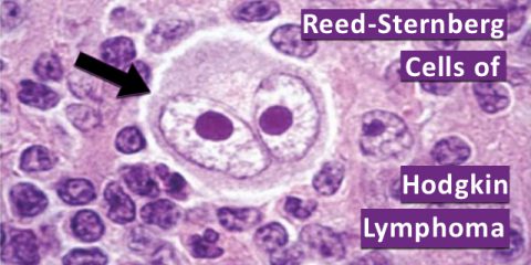
basal layer (Pagetoid spread) without dermal involvement. Hodgkin lymphoma has CD15 and CD30 positive Reed-Sternberg cells, but does not express HMB45. Squamous cell carcinoma has intradermal irregular nests or cords of atypical
squamous cells with intercellular bridges and/or keratin pearl formation, but does not express HMB45.
18. E. Sun exposure is the most important risk factor
for lentigo,
melanoma,
squamous
cell carcinoma and basal
cell carcinoma. Alcohol, cigarette and HPV are less likely associated with melanoma.
EB virus is associated with Hodgkin lymphoma.
19. C. The most important prognostic factor for invasive
melanoma is the depth of invasion, as defined by the distance between
granular layer to the deepest tumor cells.
20. B. This is an intradermal nodular growth of tumor cells
with moderate to abundant pale cytoplasm, most consistent with nodular
type invasive melanoma. Tumor cells of basal
cell carcinoma and Merkel cell carcinoma have scant cytoplasm. Melanoma
in situ is characterized by nests of atypical melanocytes at and beyond
basal layer (Pagetoid spread) without dermal involvement. Squamous cell carcinoma has intradermal irregular nests or cords of atypical
squamous cells with intercellular bridges and/or keratin pearl formation.
Back to skin
tumors
Back to contents
Comments
Post a Comment